

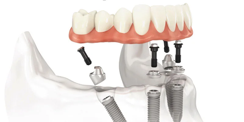
How is Implant Treatment Performed? Izmir Process Analysis and Medical Steps
Tissue losses that occur in the anatomy of the mouth are pathological conditions that bring about various changes in human physiology regarding chewing (mastication), swallowing (deglutition), and the cellular structure of the jawbone. One of the frequently used restoration methods in modern dentistry to compensate for missing teeth is the surgical placement of artificial roots made of inorganic titanium alloys into the jawbone (alveolar crest).
What is Implant Treatment and Why is it Preferred for Tooth Loss?
Implant treatment is a procedure involving the surgical placement of titanium materials that show biological compatibility (biocompatibility) with human tissue into the jawbone to restore the chewing and speaking functions of lost natural teeth. This treatment is often valued in medical practice as it does not require intervention in the enamel tissues of neighboring healthy teeth (the need for reduction) and slows down the physiological process of bone resorption at the cellular level by directly transferring chewing pressure to the bone. The artificial root placed in the jawbone integrates with bone cells (osteoblasts) at a microscopic level over a period of months, forming a mechanical and structural support unit for porcelain or zirconium crowns that will be installed on top.
The maxilla (upper jaw) and mandible (lower jaw) bones, which are parts of the human skeletal system, are dynamic tissues that maintain their shape and volume under mechanical pressures transmitted through the natural roots of teeth located within them. In cases where one or more teeth are extracted, the adjacent alveolar bone area cannot receive the physical stimulation that results from chewing forces. According to the orthopedic principle known as Wolff’s Law, the activity of osteoclast (bone-resorbing) cells increases in bone tissue deprived of mechanical stress, leading to significant tissue resorption in the area. The clinical basis of implant application is to restore this disrupted biomechanical stimulation system. The titanium alloy placed into the bone during surgery is an inorganic structure that is not rejected by the integration system.
In traditional dentistry, it is a clinical necessity to reduce the healthy teeth adjacent to the gap by cutting (milling) for bridge prostheses frequently applied for single tooth deficiencies. However, in implant operations, the enamel and dentin tissues of neighboring teeth maintain their anatomical integrity; the intervention is localized only to the area of deficiency. The hundreds of Newtons of occlusal forces that arise during the chewing process are directly transferred from the titanium screw to the alveolar bone. This transfer stimulates the cells in the bone matrix, keeping the mineralization cycle active. In Izmir Avrupadent clinics, this medical restoration is designed beyond a limited repair, aiming for the accurate digestion of the nutrients reaching the patient’s digestive system and the structural support of the jaw skeleton.
How is the Clinical Evaluation Conducted Before Implant Treatment in Izmir?
The clinical evaluation process consists of steps such as obtaining the patient’s medical and systemic history (anamnesis), examining the soft and hard tissues of the oral cavity with periodontal probes, assessing vertical occlusal relationships, and collecting radiological data. As a result of these comprehensive analyses, the physician determines the patient’s tolerance for surgical intervention, infection risk profile, and the physical dimensions of the materials to be used based on objective findings.
In the general medical management of surgical interventions, the diagnostic (diagnosis) phase planned before the operation is clinically significant, just as the procedures performed during the operation itself. This process begins with the patient’s application to the İzmir Avrupadent clinic and is deepened by completing systemic anamnesis forms. The patient’s overall health status, cardiovascular reserve, and cellular healing capacity are classified according to the American Society of Anesthesiologists (ASA) physical risk scale. When chronic diseases affecting systemic circulation or cellular oxygenation (such as diabetes, hypertension, and osteoporosis) are detected, a medical consultation for surgical intervention is requested from the medical branches the patient is being followed up in.
During the physical intraoral examination, the physician does not only focus on the missing tooth gap; they also examine the patient’s overall oral hygiene profile, the amount of plaque, and the gingival biotype (thickness of the mucosa). If the amount of keratinized mucosa surrounding the implant is insufficient, it may lead to microbial accumulation around the prosthesis in later years, so additional surgical steps such as soft tissue grafting can be included in the treatment plan. At the same time, by observing the patient’s muscle dynamics, temporomandibular joint mobility, and possible bruxism (teeth grinding) habits, the destructive effects of the vectorial forces (in Newton) that will act on the artificial roots can be calculated. Planning a surgical procedure without this data is considered contrary to medical protocols.
Why is Radiological Examination Important Before Implant Treatment?
Radiological examination maps the vertical height, horizontal width, degree of trabecular hardness (Hounsfield units), and the distance to vital organs such as nerve canals of the bone anatomy concealed beneath the mucosa. This digital analysis performed with three-dimensional dental volumetric tomography (CBCT) determines the entry angle of surgical drills into the bone, limiting structural levels of potential anatomical injury risks during the procedure.
In the oral cavity, the true form and density of the underlying bone matrix cannot be clinically understood solely by examining the surface tissues. Panoramic X-rays, traditionally used in dentistry, reflect the entire jaw structure as a two-dimensional arc, but they contain distortion (enlargement or contraction) in certain proportions (usually between 15-25%). While these two-dimensional films may be functional for detecting cysts or general root lengths, they do not physiologically reflect the thickness of the area where the implant will be placed in the anterior-posterior (bucco-lingual) direction.
To eliminate this anatomical uncertainty, Cone Beam Computed Tomography (CBCT) devices, which are modern medical technologies, are utilized. CBCT creates a digital anatomical replica of the patient by separating the jaw into millimeter slices with a low radiation dose. It is a clinical rule to leave a minimum safety distance of 2 millimeters between the ‘inferior alveolar nerve’ cord that transmits lip sensation to the brain in the lower jaw and the ‘maxillary sinus’ membrane that contains air spaces in the upper jaw. The tomographic data is transferred to software for the application of the “backward planning” process. In this concept, the ideal crown (porcelain tooth) that should be in the mouth is virtually designed; then a titanium screw is digitally placed at the most appropriate angle into the jawbone from the center of this tooth. Thanks to technological analyses at Avrupadent in Izmir, the surgical operation is carried out with mathematical navigation based on scientific data in addition to the surgeon’s experience.
What Are the Surgical Steps of Implant Treatment?The surgical steps include; under local anesthesia, cutting the mucosal tissue (gum) to expose the bone surface, gradually performing osteotomy (drilling) in the alveolar bone using micromotors that provide physiological saline cooling, placing the titanium screw into the socket with a specific torque (tightening force), and closing the gum tissue with surgical sutures.
The surgical steps include; under local anesthesia, cutting the mucosal tissue (gum) to expose the bone surface, gradually performing osteotomy (drilling) in the alveolar bone using micromotors that provide physiological saline cooling, placing the titanium screw into the socket with a specific torque (tightening force), and closing the gum tissue with surgical sutures.
On the day of the operation, clinical environmental medical asepsis and antisepsis standards are prepared to minimize the possibility of cross-infection. After the surgical area is anesthetized with local anesthetic solutions, the surgeon makes an incision on the gum tissue in the operational field with the help of a scalpel. The mucosa and periosteum (bone membrane) are raised (flap operation) to expose the underlying cortical bone layer. According to the millimetric measurements determined in digital radiological planning, a cavity is prepared on the crest with the help of progressively larger medical drills.
During this osteotomy (drilling) procedure, if the temperature of the bone tissue exceeds 47 degrees Celsius, it will lead to protein denaturation and death of bone cells (necrosis); therefore, continuous cooling of the surgical micromotors with physiological serum is a clinical necessity. When the cavity preparation is completed, a titanium implant taken out of its sterile packaging is positioned using low-speed devices or manual torque wrenches. The pressure force applied by the screw to the bone (primary stability) is typically targeted to be between 30-45 Newton-centimeters (Ncm). A cover screw is placed on the implant to protect its internal threads, and the raised gum flap is sutured with surgical threads to ensure primary wound closure. In the Izmir Avrupadent procedures, post-operative care instructions are thoroughly communicated to the patient after the operation.
| Surgical Step | Medical Procedure Performed | Clinical Function and Purpose |
|---|---|---|
| 1. Incision and Flap Elevation | Cutting the gum tissue (mucosa) with a scalpel and lifting it off the bone. | Allows for direct observation of the alveolar bone anatomy. |
| 2. Osteotomy (Cavity Preparation) | Drilling the bone at the level of liquid cooling, progressively increasing with conical drills. | Creates an anatomical socket suitable for the dimensions of the titanium material. |
| 3. Implant Placement | Insertion of the titanium screw into the prepared cavity using a torque device. | Biological groundwork is prepared by achieving mechanical compression (primary stability). |
| 4. Suturing (Stitching) Application | The lifted gum tissue is closed and sutured with surgical threads. | Supports hemostasis (control of bleeding) by ensuring the union of wound lips. |
How is Anesthesia Management Planned During Implant Treatment?
During the surgical procedure, local anesthetic pharmacological agents are injected around the major nerve bundles in the area of operation, temporarily suspending neural (nerve) conduction. Since the conduction is interrupted, the patient, who is conscious, does not feel any pain during the intervention on the bone; they only perceive the physiological vibrations of the devices and the mechanical pressure applied to the tissue.
In invasive procedures performed in the maxillofacial (jaw-face) region, pain management is provided by anesthetic solutions (such as articaine or lidocaine molecules) that stop the permeability of sodium channels in peripheral nerve fibers. The physician applies injections to anatomical points that provide the innervation (nerve supply) of the operation area (infiltration in the upper jaw, regional blocking techniques in the lower jaw). When this pharmacological effect is active, making an incision on the mucosal tissue with a scalpel or performing drilling (osteotomy) on the bone does not send a biological disruption (pain) signal to the central nervous system.
During the operation, substances such as epinephrine (adrenaline), a vasoconstrictor, are included in the solutions to prolong the effect of anesthesia on the tissue and minimize bleeding (hemostasis) in the surgical area. However, in patients with hypertension (high blood pressure) or cardiovascular rhythm disorders, solutions without adrenaline may be used based on medical assessments, as adrenaline can cause a spike in blood pressure upon entering the bloodstream. Surgical devices used during osteotomy generate some mechanical vibration in the jawbone. While local anesthesia blocks the fine nerve fibers carrying pain, it is not equally effective on the thick fibers that convey pressure and touch sensations; therefore, it is possible for the patient to feel pressure or vibration in their jaw. In clinical procedures conducted in İzmir, intravenous conscious sedation techniques are offered as a medical alternative in conjunction with anesthesiology consultation for individuals with high levels of dental phobia.
How Long Does the Bone Heating (Osseointegration) Process Take?
The osseointegration process, which is the microscopically integrating of the inorganic titanium artificial root with the alveolar bone at the cellular level, takes an average of two to three months in the lower jaw with cortical density; while in the upper jaw, which has a more spongy and aerated structure, it encompasses a biological healing period of three to six months.
During the operation, the physician achieves mechanical physical compression (primary stability) as they place the implant into the prepared socket in the bone. However, due to the fact that human bone is a living and dynamic tissue, this mechanical force decreases over the months and is replaced by cellular retention (secondary stability). The first blood clot that surfaces in the surgical trauma area creates a biological scaffold by forming fibrin strands, allowing bone cells to migrate to the area. In the following weeks, the connective tissue cells of the organism organize the wound site while bone-forming osteoblast cells settle into the porous microstructure of the titanium surface. These cells secrete calcium and phosphate minerals into the surrounding area, constructing a new, calcified (hardened) living bone matrix.
The mandible (lower jaw) is anatomically extremely hard (generally type D1 or D2) and has a high mineral density, so the processes of osteoblast proliferation and calcification turn back to mechanical stability in a shorter time. The maxilla (upper jaw), on the other hand, is adjacent to the nasal floor and sinus cavities and contains a more porous and fine trabecular structure (type D3 or D4). The mineralization of the spaces within this structure depends on the anatomical requirements and is therefore extended over a broader timeline. This waiting process is a cycle dependent on the organism’s own cellular physiology, which cannot be shortened by pharmacological agents applied externally. According to the Izmir Avrupadent protocols, periodic radiological examinations are conducted to confirm whether there is any radiolucency (darkness) in the implant-bone contact area, followed by approval for prosthesis construction.
What Additional Procedures Are Applied in Jaw Bone Insufficiency?“}If the radiological analyses indicate that the alveolar bone volume is insufficient in terms of vertical height or horizontal width, bone augmentation (graft/addition of powder) procedures are applied. In the posterior areas of the upper jaw, biological particles are filled into the void formed by lifting the sinus membrane (Sinus Lifting) to reconstruct the bone volume.
If the radiological analyses indicate that the alveolar bone volume is insufficient in terms of vertical height or horizontal width, bone augmentation (graft/addition of powder) procedures are applied. In the posterior areas of the upper jaw, biological particles are filled into the void formed by lifting the sinus membrane (Sinus Lifting) to reconstruct the bone volume.
The long-term mechanical stability of titanium material depends on the thickness of the surrounding healthy bone wall. The implant body requires at least 1.5 – 2.0 millimeters of thicker bone tissue from all sides. The passage of many years since tooth loss, along with cystic infections or severe gum diseases (periodontitis), leads to extensive volumetric defects in the cortical walls of the jawbone. In the presence of these defects, when a direct socket is opened, the grooves of the screw remain exposed; these exposed grooves under the mucosa prepare the ground for bacterial retention, creating an infection site.
To overcome these anatomical handicaps, techniques of “augmentation” (volume increase) are employed. Biomaterial graft (bone powder) particles are placed in the surgical site. These particles can come in forms from inorganic filler sources (xenograft), synthetic (alloplastic), or human donor sources (allograft). The placed graft forms a three-dimensional matrix that can migrate and calcify over the body’s own bone cells. In the posterior areas of the upper jaw where toothlessness causes the maxillary sinus membrane (Schneiderian membrane) to sag downward, it is pushed upward by special surgical instruments, and graft materials are filled into the new area formed. In clinical practices in İzmir, doctors prepare the tissue physiology according to the size of the defect by performing these advanced surgical interventions either in the same session as implantation or about six months prior to the implantation process.
| Material Type | Biological Source | Clinical Function and Reaction |
|---|---|---|
| Autogenous Graft | Living bone blocks taken from the patient’s own body (chin tip, ramus, or calvaria). | Contains living osteoblasts. Directly stimulates new bone formation (osteogenesis). |
| Xenograft | Obtained by processing animal sources such as cow, pig, or horse in the laboratory to remove proteins. | Provides a long-lasting volume that is very slow to resorb by offering a calcium scaffold. |
| Alloplastic Graft | Synthetic inorganic powders derived from hydroxyapatite or tricalcium phosphate synthesized in a laboratory environment. | Does not harbor risks of infection or antigenic reaction, provides space for cell proliferation. |
| Collagen Membrane | Thin tissue covers (barriers) derived from pig or cow pericardium. | Physically restricts rapidly proliferating gum tissue cells from entering the grafted area and disrupting osseointegration. |
How do Systemic Diseases Affect the Implant Treatment Process?
Systemic diseases such as uncontrolled diabetes, hypertension, cardiovascular disorders, or osteoporosis directly inhibit microvascular (capillary) circulation in the wound area, the coagulation mechanism, and osteoblast (cellular repair) activity. When these values are regulated to reference ranges with medical consultations, treatment can be carried out with minimized risks of inflammation.
Surgical invasive procedures performed on the tissues inside the mouth are not just localized interventions but occur in an integrated manner with the overall physiology of the organism through vascular and lymphatic systems. In patients with chronically high blood glucose (sugar) levels, advanced diabetes causes thickening and blockage (microangiopathy) in the vessel walls. This cellular vascular defect narrows the volume of blood that needs to reach the surgical area, leading to tissue remaining oxygen-deprived (hypoxic), slowing collagen synthesis at the wound site, and preventing leukocytes (defense cells) from reaching the area. Due to this biological condition, HbA1c tests, which are indicators of the patient’s blood sugar over the last three months, are requested before surgery, and a condition is required for the values to be around 7% (in clinical references).
Heart valve surgery slows the coagulation cascade in patients who regularly use anticoagulants (blood thinners) due to having a stent or atrial fibrillation (arrhythmia). In order to physiologically stop the bleeding (hemostasis) when a mucosal incision is made, medication modifications (bridging therapy) are applied with a cardiologist’s consultation. Additionally, in patients receiving intravenous bisphosphonates with a diagnosis of osteoporosis (bone loss), there is a risk of cell death (osteonecrosis) in the surgical area because the medication slows down bone formation while suppressing bone resorption. In this context, all systemic diseases of patients in the İzmir Avrupadent processes are individually medically analyzed by classifying them through the ASA (American Society of Anesthesiologists) risk scale.
How Does the Prosthetic (Crown) Phase Progress After Implant Treatment?
After the biological bone integration (osseointegration) phase is confirmed radiologically, the mucosal tissue over the implants is shaped with healing caps, and a three-dimensional copy of the jaw is obtained using intraoral (in-mouth) optical scanners or conventional systems. Zirconium or metal-supported ceramic crowns, resistant to occlusal (biting) forces, are produced in the dental laboratory and fixed to the implants with abutments.
When the cellular adaptation of the surgical phase is completed, the prosthetic rehabilitation phase is initiated, where the treatment will gain functional efficacy and aesthetic restoration will be applied. In the first step, under local anesthesia (or without the need for anesthesia if there is a thin mucosal structure), the gum (mucosa) is opened over the cover screw, and healing caps called ‘gingiva formers’ are placed. The gum mucosa takes on its physiological form around this cylindrical metal within an average of 7 to 14 days, creating a ‘pink aesthetic’ base to ensure that the crown appears as if it is emerging from the bone like a natural tooth.
After the removal of the outer tissue profile, a “measure sheet” (transfer piece) is placed inside the implant bodies. High precision silicone (elastomeric) measurement materials or computer-assisted intraoral scanners (CAD/CAM) are used to accurately transfer the jaw curves, occlusion relationships, and the amount of missing tissue to the laboratory. The aim of the prosthetic design is for the dentist to restore the patient’s Vertical Dimension of Occlusion according to skeletal muscle dynamics. In the areas of lower molar regions where chewing forces are very intense, ceramics with a chrome-cobalt metal infrastructure, which have high mechanical resilience to bending and fracture, are preferred; whereas in the anterior areas where light permeability and aesthetics are a priority, crowns with zirconium infrastructure that are close to natural enamel fracture are preferred. These produced crowns are fabricated using medical cements via an intermediary piece (abutment) attached to the implant bodies or fixed with direct screw attachment (screw system).
What Should Be Considered During the Healing Period After Implant Treatment?
To control the physiological edema that may occur after the operation, a cold compress (ice) should be applied externally to the surgical area for the first 48 hours. It is essential to adhere to the medical schedule for the antibiotics, pain relievers, and chlorhexidine-containing mouthwashes prescribed by the doctor, to consume soft foods in the initial days, and to avoid products that disrupt tissue perfusion (such as smoking) for wound healing.
Surgical incisions made to the mucosa and bone membrane inside the mouth trigger a biological repair response (inflammatory) in the body. Inflammation is the process of sending fluid, blood, and cellular elements to the damaged area to repair the affected tissue. The external manifestations of this physiological response include localized tissue swelling (edema) and, rarely, capillary bruises (ecchymosis) on the outer skin, which are commonly encountered in the early stages of wound healing. Applying cold compresses intermittently from the facial area narrows capillary diameter (vasoconstriction), limiting fluid accumulation and potential bleeding leaks.
Structural modifications in the patient’s dietary habits are required to prevent tension on the wound edges and ensure the stability of the fibrin clot in the osteotomy area. In the early days, it is important to avoid very hot beverages and acidic or spicy foods that could dissolve the clot; a puree-based diet at room temperature should be followed. The major factor that negatively affects wound physiology at the cellular level is the consumption of tobacco products. Carbon monoxide in cigarette smoke binds to hemoglobin in the blood with a higher affinity (binding desire) than oxygen, leading to decreased tissue oxygenation (hypoxia). Simultaneously, nicotine narrows the blood vessels in the surrounding tissues, reducing the volume of blood carried to the surgical site. A wound area that does not receive oxygen and defense cells struggles to close and becomes open to bacterial growth. In this context, limiting tobacco consumption to medical norms throughout the osseointegration process is strongly emphasized to patients at İzmir Avrupadent clinics.
How Should Long-Term Care for Implant Teeth Be Managed?
The long-term functional maintenance of prosthetic restorations is ensured by mechanically cleaning the microbial plaque between the prosthesis and the gum using standard fluoride toothpaste along with interdental brushes, specially designed dental floss (superfloss), and powered water devices (oral irrigators). Clinical radiological checks and professional tartar (dental calculus) cleaning performed every six months is a medical protocol.
Due to the corrosion resistance of titanium material and the inorganic structures of porcelain/zirconium crowns, it should not lead to physiological misunderstandings that imply the application does not require individual oral hygiene (no caries formation). The mucosa (gum tissue) surrounding the implant bodies and the supporting tissue of the alveolar bone is much more fragile against the microbial dental plaque in the oral cavity compared to natural tooth structures. Periodontal fibers (ligaments) in natural teeth slow the penetration of bacteria into the bone and serve as a barrier; these are not cellularly present around the inorganic titanium screw (ankylotic contact).
Due to this anatomical difference, when bacterial plaque accumulates in the fine groove (margin line) between the crown and the gum, inflammation can penetrate the bone tissue much more rapidly. Initially, soft tissue manifests as bleeding and redness, indicating peri-mucositis (inflammation of soft tissue). If mechanical hygiene is not maintained, it leads to the stage of peri-implantitis (bone resorption around the implant), causing the screw to lose its biomechanical stability. Since the accumulated inorganic dental calculus (tartar) cannot be removed with a home toothbrush, patients should periodically undergo ultrasonic detartrage procedures in a clinical setting by a doctor twice a year, and measure the depths of the gum pockets, which is the only way to protect the medical lifespan of the restoration.







