

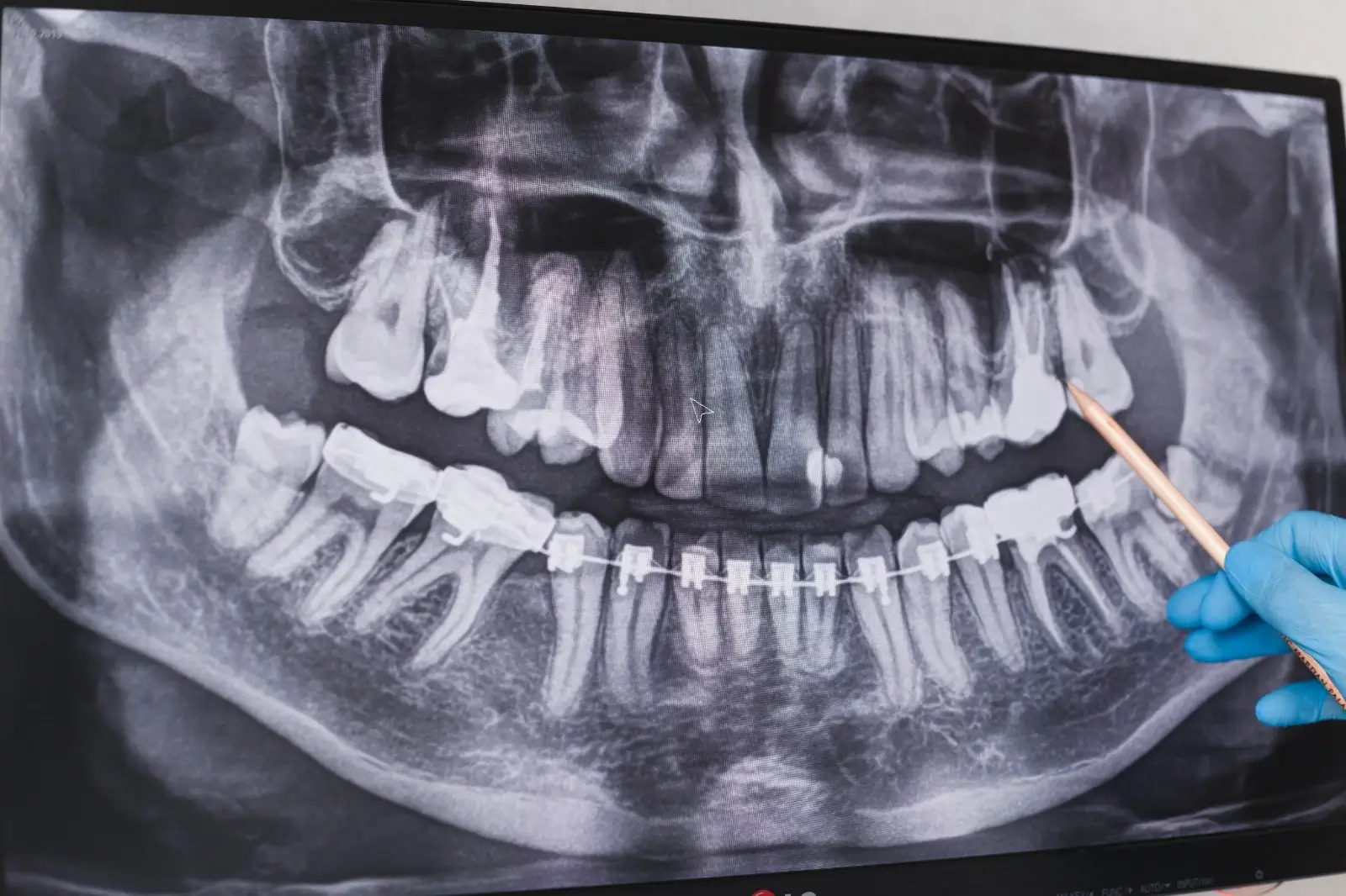
A periapical X-ray is a dental X-ray taken with a small film placed inside the mouth, showing one or two teeth in detail together with the root tip and the surrounding bone. It is one of the basic images a dentist turns to for toothache, a suspected abscess, the depth of decay and the follow-up of root canal treatment.
What is a periapical X-ray and what does it show
A periapical X-ray shows the whole tooth, crown and root tip, in a single frame. “Periapical” means the area around the root tip; the film reveals the condition of the bone and tissues there. A dark area at the root tip may point to inflammation, bone loss or a lesion. For a general explanation of dental X-rays, see our page what is a dental X-ray and how is it taken.
Which complaints call for it: pain, abscess, decay
A periapical X-ray is usually requested when there is a complaint about a single tooth. The following situations are examples:
- Constant toothache, or pain that reacts to hot and cold
- Swelling of the gum or a suspected abscess
- Suspected decay between teeth that cannot be seen by eye
- Before, during and after root canal treatment
- The need to see the root structure of a tooth before an extraction or an implant
You can find general information about pain in the article what helps toothache from a decayed tooth.
How the X-ray is taken and how long it lasts
Taking the image is brief and is usually done with a small sensor or film placed inside the mouth. The dentist or an assistant places the sensor next to the tooth to be imaged, asks you to bite down or hold it steady, and then points the X-ray device at the tooth. Staying still during the exposure improves image quality. If more than one area is needed, the procedure is repeated for each tooth.
How decay, abscess and bone loss appear on the film
On the film, decay appears as a dark area where the tooth has lost hardness; an abscess or bone loss appears as darkening around the root tip. These findings alone are not enough to make a diagnosis; the dentist evaluates the film together with the examination, your complaints and other tests. Instead of interpreting the image yourself, it is right to discuss the results with your dentist.
How it differs from a panoramic X-ray and tomography
A periapical X-ray shows a single area in detail, while a panoramic X-ray shows both jaws and all the teeth in one frame. Tomography provides a three-dimensional image and is requested when information such as bone volume is needed. So these three methods do not replace one another; the dentist chooses according to the purpose. For tomography, you can take a look at our page dental tomography (tooth tomography).
What the dentist pays attention to in children and pregnant patients
In children and pregnant patients, an X-ray is requested only when it is truly necessary and protective measures are taken. Whether an X-ray is needed for a child is assessed at the examination; for a child’s first visit, see the page a child’s first dental examination. If you are pregnant, be sure to tell your dentist before the examination.
How the results are shared with the patient
The dentist shows you the image on the screen, explains the findings and, if treatment is needed, talks through the options. In processes such as root canal treatment, the film is also used to follow how the treatment is progressing; on this subject you can look at the article signs of a failed root canal treatment. If you would like an examination for your complaint, you can make an appointment through our contact page.
Frequently asked questions
What is a periapical X-ray?
It is a dental X-ray taken with a film placed inside the mouth, showing one or two teeth in detail together with the root tip and the surrounding bone.
When is a periapical X-ray taken?
It is taken when the root of a single tooth needs to be seen, such as toothache, a suspected abscess, suspected hidden decay, during root canal treatment and before surgery.
How is a periapical film taken and how long does it take?
A small sensor or film is placed inside the mouth and the X-ray device is pointed at the tooth. The exposure is brief; staying still makes the image sharper.
Can an abscess be seen on a periapical X-ray?
A dark area around the root tip may suggest an abscess, but the dentist makes the diagnosis together with the examination and other findings.
What is the difference between a periapical and a panoramic X-ray?
A periapical X-ray shows one or two teeth in detail, while a panoramic X-ray gives a general view of both jaws in a single frame.
Why is a periapical X-ray needed in root canal treatment?
It is needed to see the structure and length of the root canal and the condition at the root tip, and to follow how the treatment is progressing.







