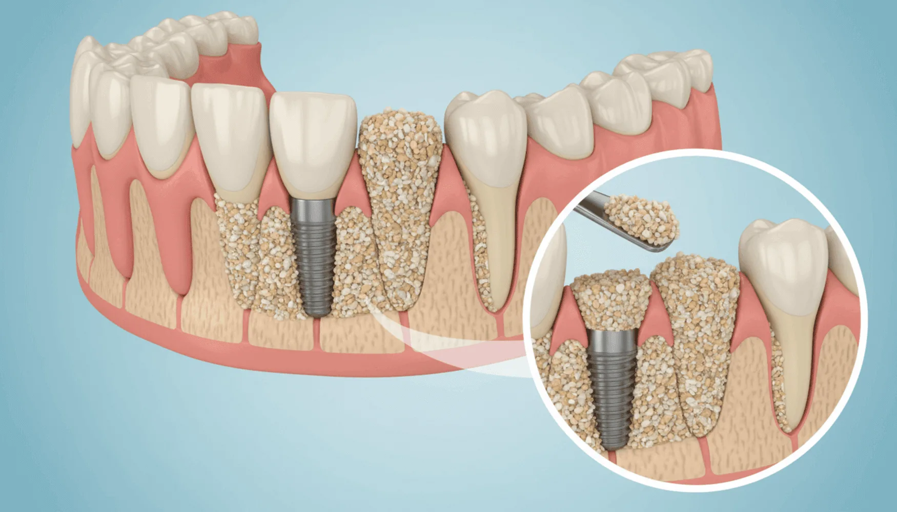
A bone graft (“bone powder”) around a tooth is done in these steps: the gum is opened, the area is cleaned, the bone graft is placed into the space, it is covered with a membrane, and the gum is closed with stitches. It is usually done under local anesthesia, and the duration varies with the condition of the bone and the area treated. Below you will find the steps in order, along with the preparation beforehand and the care afterwards.
Tests done before a bone graft
When a bone graft is planned, it must first be seen how much bone is missing. For this, a panoramic X-ray and, when needed, a three-dimensional tomography scan are requested. On the images, the height and width of the bone, the roots of the neighboring teeth and, in the upper jaw, the position of the sinus cavity are evaluated. During the tests the dentist also assesses the bone density of the area, because how much graft is used and how it is placed are planned accordingly. The dentist examines the inside of the mouth and, if there is gum disease or an infection, plans its treatment first. Be sure to mention the medications you take, any chronic conditions and your smoking at the examination; these can affect healing.
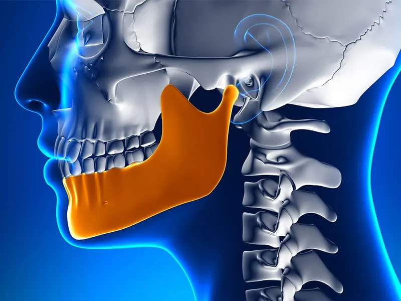
The cause of the bone loss also guides the plan. After a tooth extraction, because of gum disease, or in areas where teeth have been missing for a long time, the bone gradually recedes. You can read more about this in our article on why the jawbone matters in implant treatment: why the jawbone matters in implant treatment.
How is a bone graft done, step by step?
In general terms the procedure follows the order below. The details differ from patient to patient depending on the condition of the bone.
- Anesthesia is given. The area is numbed; during the procedure you mostly feel touch and pressure. If you feel any discomfort, it is enough to tell your dentist, and additional anesthesia is given if needed.
- The gum is opened. A small incision is made in the gum to reach the bone, and the tissue is gently lifted.
- The area is cleaned. Inflamed tissue or leftover fragments around the missing bone are removed with special instruments; a clean, bleeding surface helps the bone join with the new tissue.
- The bone graft is placed. The prepared space is filled with an appropriate amount of bone graft material and lightly packed.
- The membrane is placed. A thin membrane is laid over the graft so that it does not shift and so that the gum does not grow into the space.
- The gum is closed. The tissue is closed with stitches, which protects the area from the environment of the mouth.
- A short check is made. The dentist reviews the area and explains the care instructions both in writing and verbally.
Which anesthesia is used?
Small and medium-sized bone grafts are mostly done under local anesthesia; that is, only the area concerned is numbed and you are awake during the procedure. For procedures covering a wide area, or for patients with a high level of fear, the dentist may consider additional options such as sedation. Which method is suitable is decided at the examination according to the scope of the procedure and your health.
Why are the membrane and the closure important?
The membrane is a thin barrier that covers the graft. Because gum tissue that grows in quickly can prevent bone formation, the membrane keeps this tissue away from the graft and gives the bone time to form new tissue. Closing with stitches also protects the graft; if the closure is loose or too tight, healing can become difficult, which is why it matters that the stitches are placed properly.
What is an alloplastic graft?
An alloplastic graft is a type of synthetic bone graft made in a laboratory, not of human or animal origin. The steps of the procedure follow the same logic as with other graft types: it is placed into the space, covered with a membrane and closed with stitches. The choice between different graft types is made according to the size of the bone loss and the dentist’s plan; you can ask your dentist at the examination which material is suitable for you.
What is taken into account when a graft is placed in the upper jaw?
In the back part of the upper jaw, the sinus cavity lies directly above the bone. If the bone height is low, the sinus membrane may need to be carefully lifted and a bone graft placed beneath it. This procedure depends on seeing the position of the sinus correctly on the tomography. For more information, see our page on sinus lifting before an implant. Rules such as not perforating the membrane in the upper jaw and avoiding nosebleeds and heavy nose-blowing are also important during the healing period.
How long does the procedure take?
The duration of a bone graft operation cannot be tied to a single value. A graft in a small space can be short, while a procedure covering a wide area or the sinus region takes longer. The factors that determine the duration are the size of the bone loss, the area where it is done, and whether another procedure, such as an implant, is added in the same session. At the examination the dentist will tell you the estimated time for your own plan.
When planning the day of your appointment, it is good to allow not only for the length of the procedure but also for the preparation and a short rest afterwards. The numbness of the area can last a few hours after the procedure; during this time it is necessary to avoid hot food or drinks and to be careful not to bite your lip or cheek by accident. Not exercising heavily on the same day and resting help healing.
Preparation and care after the procedure
Before the procedure, take the medications your dentist has told you to, quit or cut down on smoking if possible, take care of your oral hygiene, and eat a light meal so that you feel alert on the day. Having someone with you is helpful, especially if sedation is being considered.
In the first days the following rules are expected to be followed:
- Do not poke the area with your tongue or fingers, and do not pull your lip back to check it.
- On the first day, avoid hot drinks and hard foods, and eat soft foods.
- Cold applied from the outside reduces swelling; use it for the time and in the way your dentist recommends.
- Use the recommended medication and, if needed, the mouthwash regularly.
- Do not skip the check-up appointments; the stitches are removed by the dentist.
Mild swelling and discomfort can be expected during this period. However, if pain increases, bleeding does not stop, or you have a fever or discharge in the area, contact the clinic without waiting. For the general course of healing, the article on the healing process after an implant may be useful.
What is done if there is not enough bone?
If the amount of bone is insufficient, the treatment plan changes: a bone graft, a sinus lift or another method may be considered. We explained how this assessment is made in our article how insufficient bone is assessed in implant treatment in İzmir. If you would like an examination for your own situation, you can choose the branch closest to you among our Çiğli, Balçova, Narlıdere, Buca, Gaziemir, Bornova, Karabağlar, Konak, Güzelbahçe, Menderes, Seferihisar and Karşıyaka branches and request an appointment through the contact page.
Frequently asked questions
How is a bone graft around a tooth done?
After anesthesia, the gum is opened, the area is cleaned, the bone graft is placed, it is covered with a membrane and the gum is closed with stitches.
How long does a bone graft operation take?
The time varies with the size of the bone loss and the area; small procedures are short, while wide ones or those in the sinus region take longer.
Which anesthesia is used for a bone graft?
Local anesthesia is mostly used; for wide procedures the dentist may consider options such as sedation.
What is an alloplastic graft?
It is a type of synthetic bone graft made in a laboratory, not of human or animal origin.
What is taken into account when a graft is placed in the upper jaw?
The position of the sinus cavity is seen on tomography; the sinus membrane is lifted carefully, and nose-blowing and nosebleeds are avoided during healing.