

Evaluation Process Prior to Dental Implant Treatment in Izmir
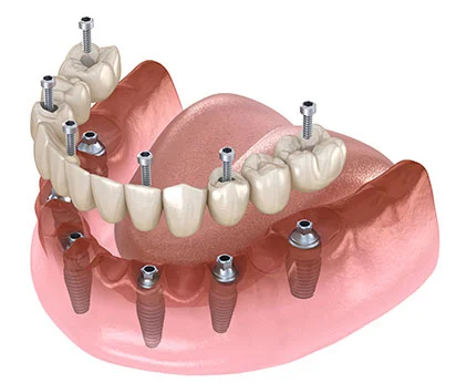
The placement of biologic materials made of titanium alloys in the jawbone, intended to compensate for missing teeth, represents a series of delicate surgical procedures in medical practice. The long-term stability of this medical procedure is based not only on the actual surgical operation but also on the comprehensive clinical analyses conducted before the operation. The evaluation process prior to dental implant treatment in Izmir is a diagnostic stage where the anatomical structure of the patient, physiological reserves, and systemic health history are mapped based on objective data. Operating in full compliance with the 2026 Ministry of Health regulations and patient information guidelines, Avrupadent addresses these diagnostic stages with a multidisciplinary approach. Advanced radiological imaging techniques, analysis of blood values, calculation of occlusal forces, and biomechanical compatibility analyses determine the clinical course of the operation.
Why is the Evaluation Process Prior to Dental Implant Treatment in Izmir a Medical Necessity?
The evaluation process prior to dental implant treatment in Izmir is a diagnostic step taken to define the anatomical boundaries of the surgical area, measure bone volume, and analyze the patient’s systemic healing capacity. Procedures performed without this evaluation carry physiological risks, such as nerve damage or medical complications.
The foundation of surgical interventions lies in anticipating the inflammatory and cellular responses that the organism will provide to the relevant intervention. The act of placing a titanium screw into the jawbone (alveolar crest) creates localized trauma. The organism responds to this trauma through clotting, cell migration, angiogenesis, and the synthesis of new bone matrix (osseointegration). For this entire biological sequence to function properly, it is clinically essential to meticulously measure the surgical site and the patient’s overall systemic structure. The individual’s existing physiological profile directly determines the timeline for cellular migration and the structural properties of the materials to be used.
Particularly, the complex anatomy of the head and neck region increases the precision of the evaluation stage. Major nerve branches, blood vessels, and the respiratory and sound resonance cavities located in the upper jaw (maxillary sinuses) pass through the jawbone and include vital boundaries that must be preserved during the operation. The pre-evaluation phase ensures that these boundaries are mapped out with millimeter precision. Avrupadent, providing medical services in İzmir, does not conduct surgical planning without obtaining diagnostic data. This meticulous process not only allows the physician to determine the surgical strategy but also forms the medical basis for all engineering calculations such as the thicknesses of the burs (drilling tools), the thread structure of the screw, and the mechanical opening of the prosthesis.
Which Radiological Imaging Techniques Are Used in Implant Evaluation?In implant surgery planning, alongside traditional two-dimensional panoramic X-rays, three-dimensional Dental Volumetric Tomography (CBCT) devices are used in current medical practice. These tomographies provide the physician with vertical and horizontal volumes of the jawbone, trabecular density, and neighboring nerve channels in millimeter sections.
In implant surgery planning, alongside traditional two-dimensional panoramic X-rays, three-dimensional Dental Volumetric Tomography (CBCT) devices are used in current medical practice. These tomographies provide the physician with vertical and horizontal volumes of the jawbone, trabecular density, and neighboring nerve channels in millimeter sections.
Radiological examination ensures the exposure of bone architecture underlying the mucosa of the oral cavity, which cannot be seen with the naked eye. Panoramic radiographs provide a general silhouette of the jawbones, tooth roots, and joints on a single plane. Pathological conditions such as cysts in the bone, buried wisdom teeth, or chronic root tip infections can be detected with these two-dimensional films. However, panoramic X-rays cannot reflect the width (bucco-lingual thickness) of the jawbone, which is a three-dimensional organ, and the images create a certain degree of structural enlargement or distortion (magnification) in the representation (between 15-25%).
To compensate for this lack of anatomical information, CBCT (Cone Beam Computed Tomography) technology is introduced. Specifically developed for dentistry, these low-radiation tomography devices acquire hundreds of millimeter slices of the jaw with a conical beam. These slices are combined in computer software to create a one-to-one digital model of the jaw. The physician can measure the mineral density of the spongy (trabecular) tissue inside the jawbone in Hounsfield Units (HU) on this 3D model. The degree of hardness of the bone (classified between D1 and D4) determines the selection of the thread design of the titanium screw to be applied and the amount of rotational torque (Newton/cm) to be applied when inserting it into the bone. In Izmir, Avrupadent processes, radiological data provide a direct data flow for the formation of surgical guides.
What Risk Areas Does Lower Jaw and Upper Jaw Anatomy Planning Include?
The ‘inferior alveolar nerve’ canal, which provides lip sensation by passing under the teeth in the lower jaw; and the maxillary sinus air spaces located in the back area of the upper jaw, which are involved in respiration, are the most critical anatomical boundaries. Planning is performed by calculating the millimetric distances to these areas.
Maxillofacial anatomy has formations differing with age, tooth loss, and individual genetic makeup. The mandibular nerve canal, which runs within the body of the mandible, is one of the most important reference points during surgery. The neurovascular bundle that passes through this canal transmits the sensation of touch from the lower lip and the tip of the chin to the brain. When preparing the implant socket, leaving at least a 2-millimeter safety margin is a clinical necessity in accordance with medical standards. The existing vertical distance between the upper crest point of the bone and the nerve canal determines the maximum length of the screw to be used.
In the upper jaw (maxilla), the maxillary sinuses neighbor the root tips of the back molars. When tooth loss occurs, the bone, deprived of the chewing force, begins to resorb, and the sinus cavities show a downward sagging (pneumatization) tendency. As a result of this sagging, there might be a bone tissue of paper-thin thickness remaining between the gum and the sinus floor. It is anatomically impossible to place an artificial root directly into this thin bone layer. In order to perform a surgical procedure in the relevant area, it is necessary first to lift the sinus floor (Sinus Lifting) and then to add biological bone grafts to the intervening space. Radiological analysis can allow for the identification of such advanced surgical needs well before the day of the operation.
How do Systemic Diseases and Medical History Shape Pre-assessments?
The patient’s existing chronic illnesses, prescribed medications, past surgeries, and allergic reactions are documented through the medical history (patient history). Hypertension, diabetes, thyroid or autoimmune disorders affect cellular healing and anesthesia tolerance, so the surgical schedule is revised according to these systemic data.
The operations performed in dental clinics are directly related to the cellular reactions of the circulatory and immune systems. The hospital’s systemic health profile influences the wound healing rate, infection resistance, and the quality of osseointegration. Therefore, the most critical part of the evaluation phase is medical interrogation (anamnesis). Patients are classified according to the physical status scale of the American Society of Anesthesiologists (ASA). Individuals in the ASA I (healthy) and ASA II (mild systemic disease) categories are included in routine clinical protocols, while priority is given to regulating the overall health of individuals in the ASA III category who have uncontrolled diseases.
For instance, the HbA1c value, which reflects the hospital’s average blood sugar level over the last three months in the presence of diabetes (diabetes mellitus), is analyzed. High blood sugar reduces the amount of oxygen reaching the wound tissue by impairing the capillary structure (microangiopathy). A decrease in oxygenation slows the activity of osteoblasts (bone-forming cells) and increases the risk of infection. The dentists at Europadent, based in Izmir, determine the appropriate timing for surgery by requesting up-to-date laboratory tests from diabetic patients or consulting with endocrinology specialists. Similarly, specific antibiotic protocols are planned for infection prophylaxis in patients with a history of organ transplantation or those using immunosuppressive (immune-suppressive) medications due to rheumatoid arthritis.
| Systemic Condition / Disease | Possible Biological Effect on Surgery | Clinical Evaluation and Action Plan |
|---|---|---|
| Uncontrolled Diabetes (Type 1 / Type 2) | Reduces collagen synthesis, suppresses cellular defense, delays wound healing. | The operation requires measuring fasting/postprandial blood sugar. The HbA1c level is expected to be around the clinical reference (%7). |
| Hypertension (High Blood Pressure) | Surgical stress and the contents of local anesthetics can lead to acute increases in blood pressure. | Choosing an anesthetic solution that does not contain a vasoconstrictor. Monitoring during the procedure. |
| Radiotherapy History (Head-Neck) | The treatment of the radiation weakens the tissue by reducing the blood vessels in the jawbone (vascularization). | Consultation with a radiation oncologist. The duration of radiation treatment is examined to assess the risk of bone necrosis. |
| Osteoporosis (Bone Loss) | Drugs from the bisphosphonate group slow down the bone remodeling cycle. | The duration of medication use, dosage, and administration route (oral/intravenous) are examined to create a medical suitability map. |
How is the Pre-Evaluation Conducted in Patients Using Anticoagulant Medication?
In individuals using anticoagulants (anticoagulant/antiplatelet) due to cardiovascular disorders or arrhythmias, the coagulation mechanism has not been suppressed. To ensure bleeding control after surgical incision, INR values are examined by contacting the cardiologist the patient is being followed by, and medication adjustments are made.
Intraoral surgical procedures create a vascular (vascular) injury in the mucosal and bony tissue. In a healthy physiology, this injury results in the accumulation of platelets (blood cells) in that area and the formation of fibrin threads, which controls the bleeding within seconds (hemostasis). Clot formation is also the initial stage of the osseointegration phase around the titanium screw. The more stable the clot is, the more organized the migration of bone-forming cells to that area becomes.
Patients who have undergone heart valve surgery, have had stents placed, or have a history of atrial fibrillation (arrhythmia) use pharmacological agents that slow down clotting. Making an incision in the gum while under the influence of these agents may lead to difficult-to-stop prolonged bleeding. In the pre-evaluation phase, the molecular structure of the medication used by the patient (whether it is an aspirin derivative, a warfarin derivative, or a new generation molecule) is determined. According to the medical protocols at İzmir Avrupadent, it is not requested for the patient to discontinue their medication on their own initiative. A consultation form written to the relevant cardiology or internal medicine specialist is sent, and a suitable “bridging” therapy (for example, transitioning to oral tablets or short-acting subcutaneous injections) is ensured under medical supervision.
What Factors Affect the Decision to Operate on Jawbone Volume and Density?
The diameter and length measurements of the implant are determined according to the physical volume of the bone tissue in which it will be placed. There must be a healthy bone wall of at least 1.5 millimeters thickness around the threaded artificial root; the vertical height of the bone must also be sufficient not to contact anatomical cavities.
In clinical evaluations, the anatomical form of the alveolar crest (the bone cavity where the teeth are located) is a concrete piece of information that determines the course of surgery. The alveolar bone, deprived of biomechanical stimulation after tooth extraction, is physiologically resorbed (absorbed) by the organism. The resorption process primarily narrows the horizontal (buccal-lingual) width of the bone, and over the years, it causes vertical (occluso-apical) height loss. If the bone volume in tomographic sections is insufficient even for the narrowest diameter screw to be used, standard implantation procedures cannot be applied.
Bone density (quality) is another critical component. The anterior region of the lower jaw has a very hard and compact (D1 or D2 type) bone structure, while the posterior region of the upper jaw is in a spongy (D4 type) form that contains large air spaces. While primary stability (initial mechanical retention) is very high in hard bone, the screw may struggle to compress the bone tissue in soft bone. The concerned physician plans specific surgical channel opening techniques based on these differences in density observed on the tomography. For example, while creating a socket in spongy bone, instead of removing the bone externally, the use of handheld instruments that compress towards the walls (osteotomes) may be preferred.
What Reconstructive Options Are Considered When Bone Insufficiency Is Detected?If it is determined that the bone volume is limited in radiological examinations, “bone augmentation” is added to the surgical plan. Advanced surgical alternatives such as the expansion of the alveolar crest using biomaterial bone grafts (powders) and barrier membranes or the elevation of sinus cavities (sinus lifting) are activated.
If it is determined that the bone volume is limited in radiological examinations, “bone augmentation” is added to the surgical plan. Advanced surgical alternatives such as the expansion of the alveolar crest using biomaterial bone grafts (powders) and barrier membranes or the elevation of sinus cavities (sinus lifting) are activated.
In medical practice, tissue engineering products are used to replace tissue deficiencies. In cases where the jawbone is too thin to accommodate an implant (in the form of a knife-edge), the surgical area is supported with biological graft particles. These particles can be sourced from inorganic block materials (xenograft), synthetic (alloplastic), or human donor sources (allograft). The materials applied create a three-dimensional scaffold that the body’s own bone cells can migrate onto and mineralize.
In clinical procedures carried out in Izmir, Avrupadent performs single-stage or two-stage reconstructive planning based on the size of the anatomical defect of the patient. If the bone deficiency is minor (small), bone powder application can be performed in the same session as the implant placement. However, if the defect is major (large) and the screw cannot provide stability, only the bone augmentation surgery is initially performed. A biological healing period of 4 to 6 months is followed for the graft material to integrate with the patient’s living bone (angiogenesis). Once the bone volume reaches the targeted width, the placement of the artificial root is carried out.
| Type of Biomaterial | Source / Production | Clinical Behavior and Healing Process |
|---|---|---|
| Autogenous Bone | Living tissue taken from a different region of the patient’s jaw (ramus or mandible tip). | Contains living osteoblasts. Directly induces new bone formation, the material with the highest biological compatibility. |
| Xenograft | Inorganic matrix obtained from animal sources like cattle or pigs, processed in the laboratory. | The re-absorption process is very slow. It provides a volume-protective structure for many years. |
| Barrier Membrane | Thin, tissue-covering medical barriers made of collagen (resorbable) or PTFE (non-resorbable). | Physically limits the invasion of rapidly proliferating gum cells into the graft area, preventing bone formation disruption. |
How is the Gum and Soft Tissue Biotype Assessment Included?
The long-term health of the implant relies on the thickness of the surrounding soft tissue (keratinized mucosa). In areas with thinner biotypes, fragile and inadequate gum tissue, soft tissue grafting is added to the planning to prevent food accumulation and bacterial infiltration around the prosthesis.
Although the primary focus in implant surgery planning is usually hard tissue (bone), the clinical importance of the mucosa (gum) biotype is equally critical. The oral cavity is a humid ecosystem that harbors hundreds of different microbial species. The junction point of the titanium screw with the jawbone is isolated from the external environment by the gum. Periodontal fiber attachments around natural tooth roots, which slow down inflammation reaching the bone, do not form on the inorganic implant surface. Therefore, the presence of a thick, immobile, fibrous band of “soft tissue” tightly surrounding the prosthetic neck is the only biological barrier against infections (peri-implantitis).
During the clinical examination, the doctor analyzes the thickness and mobility of the gum tissue using a periodontal probe (a millimetric measuring device). In areas where there is a previous (transparent) biotype or where the frenum continuously pulls the gum tissue, there is a possibility of gum recession and the gray reflection of metal under porcelain occurring in later stages. To prevent this aesthetic and medical complication, soft tissue surgery can be planned during the preliminary assessment stage. In the processes of İzmir Avrupadent, if deemed necessary, a millimetric thick connective tissue graft can be taken from the patient’s palate region and transferred to thicken the mucosa around the implant.
How are Chewing Forces and Biomechanical Parameters Calculated?
The missing tooth area of the patient, occlusal (closure) relationships, and any possible tooth grinding (bruxism) habits are examined by the doctor. By calculating the vertical and horizontal forces that will occur during chewing in Newton, the diameter, length, and material strength of the prosthetic superstructure to be used are determined.
The primary function of the prosthetic restoration and the artificial root beneath it is to smoothly transmit the intense mechanical force generated by the human jaw muscles (masseter and temporal muscles) to the bone. The area of the back molar teeth is where food is chewed, and during a single chewing cycle, it can be subjected to vertical forces ranging from 500 to 800 Newtons. The anterior incisor teeth, on the other hand, generate force primarily from horizontal directions since they are more involved in slicing.
In the reassessment phase, the mesio-distal (front-back) distance of the missing tooth gap is measured with calipers or digital scanners. To prevent the force from concentrating on a single point in the posterior regions, wider diameter (for example, 4.5 mm – 5.0 mm) implant bodies are preferred according to anatomical allowances. If the patient is diagnosed with involuntary teeth grinding or clenching (bruxism) during sleep, the treatment plan is revised to reduce the harmful and excessive loads on the prosthetic system. The biting surfaces of zirconium or metal-supported ceramic crowns are designed in the laboratory to direct the forces vertically to the long axis of titanium. This biomechanical compatibility clinically reduces the breakage or loosening of mechanical parts (abutment screws).
What Risk Factors Indicate During the Evaluation Phase of Smoking Consumption?
The consumption of tobacco products is a factor that directly inhibits cellular circulation (microcirculation) and tissue oxygenation. Intense smoking identified in the anamnesis phase recalibrates surgical treatment decisions due to risks of delayed wound healing, failure of bone grafts, and the slowing of bone cell activation.
In intraoral surgical operations, cellular wound healing progresses in proportion to the quality of local blood flow. Nicotine, the main pharmacological agent in tobacco smoke, causes the constriction of peripheral capillaries in systemic circulation (vasoconstriction). The narrowing of the vessel lumen significantly reduces the blood volume reaching the operated bone and mucosal tissues. Simultaneously, the carbon monoxide molecule in the smoke binds to hemoglobin that carries oxygen in the blood and blocks the amount of oxygen reaching the tissues (hypoxia).
The ability of the blood clot surrounding the implant site to organize a new vascular network (angiogenesis) and generate new bone (osteogenesis) depends on the presence of oxygen. In cases of ischemia (insufficient blood supply), the clot cannot nourish itself and may become necrotic, turning into an infection site. Additionally, smoking can suppress the immunoglobulins (protective antibodies) in saliva, allowing opportunistic bacteria in the oral flora to proliferate. In the examinations conducted at Avrupadent, located in Izmir, these biological interactions are conveyed to patients with clinical data. According to medical protocols, it is recommended to stop using tobacco at least a few weeks before the operation day and to significantly limit it during the healing period, to ensure that physiological wound healing is not impaired.
How Does Surgical Guide Planning Work at Izmir Avrupadent?
Using the 3D tomography data obtained from the patient, optical scanning data of the oral cavity is overlaid in a computer environment. ‘Backward planning’ is performed to calculate the most suitable bone angle for the position of the prosthesis to be placed, and medical plates that physically dictate this angle to the surgeon during the operation are produced with 3D printers.
In modern dental surgery, digital workflows (CAD/CAM technologies) are used to minimize millimeter deviations that may arise from human factors. The concept of ‘backward planning’ reverses classical surgical approaches. In the traditional method, implants are placed where the bone is located, and the prosthesis is attempted to be adapted to that angle. In digital planning, however, the process works in reverse; firstly, the ideal porcelain tooth is virtually designed on the computer screen according to the patient’s facial physiognomy and opposing jaw relationships.
Afterwards, a virtual implant is positioned precisely in the center of this ideal prosthesis to transmit the chewing force along the most uniform axis to the bone. The physician checks the distance of this virtual screw to nerves, vessels, or adjacent tooth roots in millimeters on the tomography slices. Once the planning is approved, this digital data is sent to 3D printers to produce a surgical guide (template) made of acrylic resin. During the operation, this guide, which sits on the patient’s jaw, has metal sleeves that direct the drilling burs only at the planned opening and millimeter depth. This technological preparation phase allows the socket preparation to be performed safely within medical boundaries, even without opening the flap (soft tissue incision) (suture-free operation).







