Do implants look and feel like natural teeth?
Prosthetic restorations that replace missing teeth have evolved from being merely mechanical chewing tools in modern dental technology to becoming an anatomical structure that interacts with biological tissues. The visible components, made from ceramic or zirconium on titanium materials that assume the function of the dental root, are designed to support the normal functioning of the jaw complex. The question of whether implants look and feel like natural teeth is one of the most frequently researched topics for individuals planning treatment who want to restore chewing mechanics and maintain aesthetic integrity.
Do implants look and feel like natural teeth?
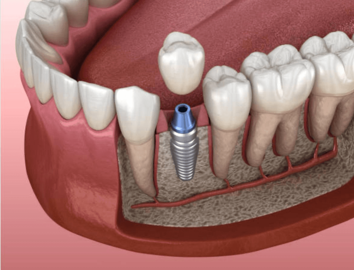
In dental practices, the concept of “naturalness” consists of two fundamental components: aesthetic illusion and functional adaptation. The aesthetic aspect is based on the principle that the prosthesis should not be distinguishable from the neighboring anatomical structures when viewed from the outside. The functional aspect is that the patient should not feel the presence of a foreign object in their mouth while speaking, swallowing, and chewing. The titanium screws positioned within the bone, along with the applied superstructure (crown), are produced according to medical standards that will meet these two expectations to a large extent.
When titanium material fuses with the jawbone at a microscopic level (osteointegration), the artificial root is mechanically anchored like our own roots. This stability eliminates the feeling of movement or vibration experienced with removable prosthetics (dentures), providing the individual with a stable and usual comfort zone. Once the healing period is over, the system becomes a part of the organism.
To what extent do anatomical teeth resemble the chewing sensation?
Our own anatomical teeth are held by microscopic connective tissue fibers known as the “periodontal ligament”, which serve as a cushion within the jawbone. These fibers allow the tooth to flex slightly within the bone when a hard object is bitten, absorbing pressure. However, titanium screws used in jaw surgery lack this connective tissue; the material bonds directly to the bone through ankylosis (fusion).
From a physiological perspective, the main sensation felt by the patient during chewing is the pressure and stability transmitted to the brain during the fragmentation of food. The titanium structure allows pressure waves from the bone to be communicated to the nerve endings (proprioceptors), enabling the patient to perceive their chewing force. Therefore, while there is a lack of connective tissue at the cellular level, the chewing sensation felt at the macro level is very close to that of a natural tooth.
How are zirconia and porcelain materials designed for aesthetic compatibility?
The way a prosthesis looks as if it belongs to the anatomical structure when viewed from the outside is related to the light absorption and reflection capacity of the surface tissue. Human teeth consist of layers with different light transmittance, namely enamel and dentin. The tips of the teeth are more translucent, while the color intensifies towards the boundary with the gums. Prosthetics produced in a laboratory environment are obtained by firing ceramic powders in layers that mimic these anatomical color transitions.
| Material Type | Light Transmittance | Area of Use and Aesthetic Compatibility |
|---|---|---|
| Zirconium Framework Porcelain | Transmits a high amount of light, is translucent. | In front area tooth deficiencies along the gum line. Exhibits optical properties closest to natural enamel. |
| Metal Framework Porcelain | Does not transmit light due to the metal skeleton, is matte (opaque). | In back area (molar) teeth. Shows high resistance to chewing forces but can create a thin gray reflection over time at the gum line. |
| Full Ceramic (E-max, etc.) | Has maximum light transmittance. | In single-unit anterior restorations. Supports visual illusion due to its non-opacity. |
What Medical Procedures Are Followed for Natural Appearance in Clinics Like Avrupadent?
The fundamental rule of medical aesthetics is to create a design compatible with the biological environment. Merely producing a white and flawless tooth is not sufficient to achieve a natural look. In institutional structures where oral and maxillofacial surgery and prosthetic expertise converge, a medical protocol known as “backward planning” is followed. In this method, the digital design of the prosthesis suitable for the patient’s facial contour is completed before the surgical phase.
The process of directing the surgery according to the prosthetic expectations includes the following steps:
-
\t
- Facial Analysis: The smile line, lip support, and facial symmetry are recorded in photographic studios or scanners.
- Digital Trial (Mock-up): The shape of the new teeth is presented to the patient in a digital environment for approval.
- Surgical Guide Production: Guides are produced on 3D printers to ensure that the titanium screw is placed perfectly into the axis that will carry the approved prosthesis.
\t
\t
Does Titanium Material Create Any Foreign Body Sensation in the Body?
The human body exhibits an inflammatory reaction to most metals or foreign substances that enter the tissues, attempting to expel that substance. However, thanks to the oxide layer formed on its surface, titanium is one of the medical elements that does not create a rejection reaction in the adhesion system. This biocompatibility allows bone cells (osteoblasts) to envelop the screw surface as if it were the body’s own mineral.
In the first weeks following surgical intervention, it is common to feel a slight fullness in the area due to swelling and tissue repair processes. However, as the stage of biological integration (osteointegration) is completed, the brain maps that material in the area as part of the body (neuroadaptation). After this point, the perception of foreignness disappears during physiological movements like chewing, speaking, and swallowing.
How Does Gum Profile Design (Pink Aesthetics) Integrate with Implants?
The other half that is as important as having white teeth (white aesthetics) is the healthy gum tissue surrounding the teeth (pink aesthetics). Even if a perfectly designed zirconium crown is placed on a titanium root, if there are asymmetries or black triangular gaps at the gum line, the naturalness is lost. Dentists manipulate this tissue in surgical and prosthetic stages to create a natural emergence profile.
- Temporary Crown Applications: Temporary teeth used during the healing process help reshape the structures known as papillae (the triangular gum tissue between two teeth) by applying pressure to the gum.
- Gum Grafts: In cases where tissue thickness is insufficient, a thin tissue (connective tissue graft) taken from another area of the palate is transferred to the relevant area to thicken the gum. This procedure helps limit potential gray metal reflections in the future.
What is the Physiological Role of Titanium Roots in Preserving Jawbone Volume?
The rule that “unused organs atrophy” is also valid for jawbones. When a tooth is extracted, since no chewing force is applied to the bone tissue in that area, the body perceives that bone as unnecessary tissue and starts to slowly resorb it. This volume loss lays the groundwork for the collapse of lip support in later years, leading to an aged appearance and speech disorders.
While traditional bridge prosthetics cover the edentulous area, they do not transmit pressure to the underlying bone, leading to continued bone resorption over the years. However, materials anchored to the jawbone directly transfer the chewing load to the bone by mimicking the mechanics of a natural tooth root. When bone cells sense this pressure, they remain active and maintain their structural integrity. This physiological process helps the patient preserve the natural anatomy of the jaw for many years.
How Do Prosthetics Used During Speech (Phonation) Interact?
The act of speaking is realized when air flows from the lungs through the vocal cords and then transforms into specific letters through the coordinated movements of the tongue, lips, and teeth. Particularly, for the accurate pronunciation of consonants like ‘S’, ‘T’, ‘D’, ‘N’, and ‘F’, the tongue must contact the back parts of the front teeth at a specific distance. Tooth loss or mismatched prosthetics disrupt this mechanism, causing phonation (sound production) problems.
The design of prosthetics applied in clinics located in Izmir aims for functionality, not only considering aesthetic factors but also ensuring that thickness and contour do not disrupt airflow. The volume of crowns is maintained in line with the thickness of neighboring natural teeth, allowing for sound production within the possible limits without feeling an impact from a foreign body.
How Long Does It Take for Adaptation to Real Tooth Sensation to Settle After Treatment?
The integration of a medical device into the entire body is a process that occurs not only at the cellular level but also at the neural (neurological) level. When the crown (dental covering) is first placed in the mouth, the tongue tends to frequently probe the surface of the new tooth due to a prolonged absence in that area. Similarly, the cheek mucosa and lips also work to adapt to this new volume.
| Timeline | Physiological Condition Felt by the Patient | Clinical Expectations |
|---|---|---|
| First 1 – 3 Days | Feeling of unfamiliarity, continuous contact of the tongue with the prosthesis, slight increase in gag reflex. | The brain analyzes the new physical structure in the oral environment and identifies boundaries. |
| End of 1st Week | Reduction (if any) of minimal slurring in speech, adaptation of the inside of the cheeks. | The soft tissues complete their stable arrangement around the prosthesis. |
| 2 – 4 Weeks Later | Subconscious acceptance, normal distribution of chewing force. | Neuro-adaptation is complete, and the patient begins to perceive their prosthesis as a part of their own body. |
Is there a change in the color or shape of the implant over time?
Natural teeth can become discolored over time due to their porous (tubular) structure at the microscopic level, which allows liquids to seep in. However, dental ceramics that are fired at high temperatures (typically 1000°C) and glazed achieve a glassy and non-porous surface. This glassy surface does not allow coloring agents to penetrate the material. Any plaque that may accumulate on the surface can be easily removed with a simple clinical polishing procedure.
However, even if the material itself does not degrade, biological changes in the mouth can indirectly affect the aesthetics of the prosthesis. Periodontal issues arising from general gum recession associated with aging or inadequate tooth brushing can disrupt the harmony between the prosthesis and the gum. At this point, it is imperative to ensure daily oral hygiene and to regularly undergo professional dental check-ups at least every six months.