How is the Implant Treatment Process Carried Out in Izmir?
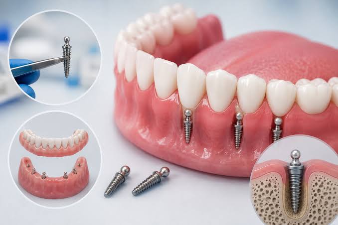
The implant treatment process in Izmir starts with a detailed clinical and radiological examination, followed by a surgical stage where medical titanium roots are placed into the jawbone. This is a multi-stage medical procedure that culminates in the fitting of permanent prosthetics prepared in the lab after waiting for the integration of bone cells with the material (osseointegration). This process is tailored specifically to the patient’s anatomical structure and biological healing speed.
This medical method applied to compensate for tooth deficiencies is a biological process based on the cellular repair capacity of the human body, rather than a standard mounting procedure. Each step of the treatment involves the protection of surrounding tissues (gum, bone, nerve pathways) and aims to restore the chewing system to a functional state. In the clinical operations within the Izmir region, the patient’s oral condition is not treated as an isolated area, but as an integrated whole with the jaw joint and overall systemic health. Dividing the process into stages is a fundamental necessity to ensure the complete implementation of medical safety protocols and to objectively verify recovery periods with tests.
How is the Initial Examination Phase Realized at the Izmir Avrupadent Clinics?
In the initial examination phase at the Izmir Avrupadent clinics, specialist doctors listen to the patient’s general health history, clinically examine the soft and hard tissues in the mouth, and volumetrically map the jawbone using three-dimensional tomography (CBCT). The objective radiological data obtained at this stage forms the basis for personalized surgical planning.
The initial evaluation is the most critical period in terms of the course of treatment. Not only the area of the missing tooth is examined, but also the root positions of adjacent teeth, the closure relationship with opposing teeth (occlusion), and oral hygiene habits are reported in detail. In the processes at Avrupadent, the reasons for procedures to be performed on the patient are clearly explained through anatomical models and digital scans. If there is an existing gum infection (periodontitis) in the patient’s mouth or untreated deep caries, the cleaning of these microbial foci is planned first. Performing a surgical procedure in an infection-free, sterile oral environment is a prerequisite for a healthy progression of the process.
| Evaluation Area | Contents and Purpose of Medical Examination |
|---|---|
| Soft Tissue (Gum) Condition | The thickness (biotype) of the gum and the inflammation status are examined to predict the wound healing capacity after surgery. |
| Closure Dynamics (Occlusion) | The contact points of the existing teeth are analyzed to calculate the chewing load that the future prosthesis will be exposed to. |
| Oral Hygiene Index | The individual’s daily brushing and care routines are evaluated; in cases of insufficient hygiene, motivation and education are provided first. |
| Adjacent Tooth Anatomy | The root resorptions of the teeth adjacent to the missing area and whether they have tilted towards the gap are determined. |
How is the Suitability of the Jawbone for Treatment Measured Radiologically?
The suitability of the jawbone for treatment is objectively determined by measuring the bone width, vertical height, and the density of the internal trabeculae in millimeters with the help of three-dimensional dental volumetric tomography (CBCT). In cases where traditional two-dimensional films are insufficient, these digital scans help identify critical anatomical boundaries such as nerve canals and sinus cavities.
The diameter and length of the titanium material to be applied must be selected in such a way that it perfectly fits the current bone volume of the patient, rather than standard measurements. The tomography device displays all cross-sections (coronal, sagittal, axial) of the jawbone on the computer screen. The physician measures how far the artificial root to be placed in the virtual environment is from the mandibular nerve, which provides lip sensation in the lower jaw, or whether it touches the maxillary sinus membrane in the upper jaw. This digital planning (simulation) allows the procedure to be completed within the medical safety boundaries by eliminating anatomical surprises that may be encountered during surgery.
Why is Systemic Health Status Questioned Before Surgical Planning?
Questioning the systemic health status before surgical planning is a medical necessity, as chronic diseases like diabetes, hypertension, heart disorders, or osteoporosis directly affect tissue healing speed, coagulation profile, and bone metabolism. By evaluating the regular medications used by the patient and their medical history, all safety protocols during the procedure are formulated based on these physiological data.
The human body consists of systems that work together in an integrated manner. In dentistry, a local intervention is related to the overall health of the body through the circulatory system. For instance, in a diabetic patient with uncontrolled (high HbA1c value) blood sugar, the microvascular structure (capillaries) is disrupted, which slows down the healing of the surgical area and makes the region prone to infection. In patients using anticoagulant medications, the management of bleeding during the procedure should be adjusted under the supervision of a medical doctor. In this regard, obtaining an accurate medical history is the cornerstone of the treatment.
- Osteoporosis Treatments: The use of bisphosphonate group medications that increase bone density can significantly impact bone healing after jaw surgery.
- Cardiovascular Diseases: A history of hypertension requires special selection of the content (epinephrine level) of the local anesthetic solution to be used during surgery.
- Autoimmune Disorders: Disorders or medications that suppress the immune system can reduce the tissues’ defense against bacterial agents.
What Steps Does the Surgical (Infrastructure) Phase of Treatment Involve?
The surgical phase of the treatment includes the local anesthesia of the relevant area, accessing the jawbone by properly retracting the gum tissue, preparing the anatomical sockets at the right opening and depth according to radiological planning, and placing biocompatible titanium material into the bone. The procedure is completed by adhering strictly to medical guidelines in a sterile clinical environment.
The surgical stage is the phase where the biomechanical basis of the procedure has been established. During this procedure performed under local anesthesia, the tissue in the area becomes numb. The surgeon gradually expands the bone using medical drills (special tools) without overheating it, at the equivalent of ample physiological saline cooling. Preserving the vitality of the bone (preventing necrosis) is the most important technique that makes the procedure successful. After the material is placed, the gums are closed with tissue-compatible stitches and left to heal. About a week later, the stitches are removed, and a tissue check is performed, marking the end of the first acute phase of the surgical stage.
| Step of the Procedure | Details of the Medical Application |
|---|---|
| Local Anesthesia and Isolation | Achieving numbness in the area through anesthesia and ensuring sterilization conditions (isolation). |
| Flap (Gum) Elevation | Creating a non-traumatic (without causing damage) separation of the gum tissue to reach the surface of the jawbone. |
| Osteotomy (Preparation of the Socket) | Opening a socket suitable for titanium material with speed and cooling systems that will protect the vitality of the bone structure. |
| Placement of the Material | Positioning of medical titanium in the bone with a specified torque (compressive force) value and suturing the gum. |
What Additional Procedures are Involved in Jawbone Insufficiency?
When it is determined that the volume of the jawbone is insufficient in radiological measurements, additional surgical procedures such as the placement of bone powder (graft) in the horizontal or vertical direction, and sinus floor elevation (sinus lifting) in the posterior regions of the upper jaw come into play. These procedures aim to reconstruct the biomechanical support tissue and volume necessary for the titanium roots to be placed.
After tooth loss, a physiological bone resorption process begins in the jaw areas that have been empty for a long time. If there is not enough healthy bone tissue around the structure to be placed (typically at least 1-2 mm thick in all directions), chewing forces can dislocate this structure in a short time. By using biocompatible bone grafts and special barrier membranes, cellular activity in the area is encouraged. The body gradually surrounds this added material with its own cells, transforming it back into original bone. However, this repair process requires an additional healing period of approximately 4 to 6 months to the treatment timeline.
How Long Does Biological Integration (Osseointegration) Take in Implantology Procedures?
Implantology biological integration (osseointegration) process takes an average of 2 to 3 months in the lower jaw and 3 to 6 months in the upper jaw, depending on the patient’s bone density and any additional surgical procedures (such as bone powder). This timeframe is the physiological waiting phase required for bone cells to completely cover the titanium surface and gain cellular resistance against functional chewing forces.
Bone healing is a cellular (microscopic) reaction that cannot be observed externally. Osteoblasts, the bone-forming cells in the human body, migrate to the specially purified surface of medical titanium used in the laboratory and colonize there. If a permanent denture is placed on the material and chewing force is applied before this colonization is complete, that is, before the connections have hardened sufficiently, the resulting micro-mobility disrupts the osseointegration and allows fibrous (soft) connective tissue to take its place. Adhering to the healing period in medical procedures is one of the structural components of the application.
How Does Temporary Tooth Application Contribute to the Healing Process?
The temporary tooth application protects the aesthetic integrity by preventing the lip profile from collapsing, especially in cases of tooth loss in the anterior region, while waiting for bone healing. It anatomically guides the gum tissue to shape itself appropriately for the permanent prosthesis. Additionally, it significantly contributes to the patient’s social life during the months-long waiting period by supporting their speaking (phonetic) functions.
In cases where aesthetic concerns are high, being toothless can be psychologically challenging for patients. These temporary prosthetics, made from lightweight acrylic or similar materials, are specifically adjusted not to exert pressure on the healing surgical area (they will remain in passive contact). Patients should be informed that the temporary teeth are placed purely for aesthetic and tissue shaping purposes, rather than for chewing. A fundamental patient rule is that hard or tearable foods should not be bitten with these teeth, as it is essential for the health of the underlying bone healing process.
How to Proceed with Prosthetic (Superstructure) Process Once Bone Healing is Complete?
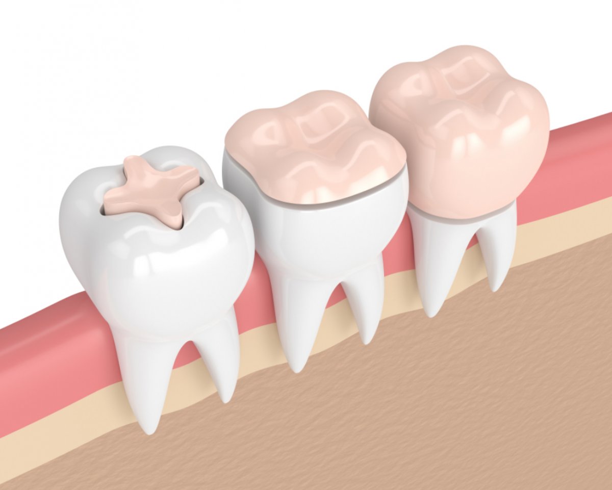
Once bone healing is confirmed through radiological and clinical tests, the healing caps on the titanium roots are removed, and 3D impressions are taken inside the mouth using digital scanners or precision silicone materials, followed by the prosthetic (superstructure) process. Zirconia or metal-supported porcelain crowns prepared in the laboratory are fixed to the substructure after ensuring a balanced occlusion relationship through try-in.
The prosthetic phase is the final step where aesthetics and function are constructed on the surgical foundation. Thanks to the impression procedure, the patient’s jaw closure, the position of adjacent teeth, and the gum’s formed “emergence profile” are accurately communicated to the dental technicians. The prepared tooth framework (substructure try-in) is tested in the mouth; the relationship with surrounding tissues, the compatibility with the patient’s facial contour, and lip support are examined. Then, the color of the porcelain is determined (to match the adjacent teeth), and the final firing process is carried out. When the dental restoration is fixed in the oral cavity, it is ensured that the chewing balance is evenly distributed.
| Process Step | Clinical Detail of the Procedure |
|---|---|
| Tissue Shaping | Use of healing caps (gingiva former) to ensure the gum takes a natural shape before the final denture is installed. |
| Precise Impression Taking | Copying the mouth using digital (scanner) or conventional silicone methods with transfer pieces. |
| Metal/Zirconia Try-in | Testing the compatibility of the inner skeleton of the tooth within the mouth, the mechanical support, and the seating on the gum. |
| Permanent Fixation | The crowns that pass the aesthetic, color, and occlusion tests are connected to the infrastructure using special medical screws or resin (constructor). |
How are Routine Post-Treatment Check-Ups Organized at the İzmir Location?
At the clinic in İzmir, routine check-ups after treatment are scheduled for the first week, the first month, and the sixth month during the first year in order to closely monitor the tissue compatibility and personal hygiene habits of the patient. In the following years, periodic examinations every six months or annually are organized to radiologically assess the health of the gum tissue around the prosthesis, bone level, and occlusal balance.
The end of treatment does not mean the termination of the doctor-patient relationship. The long-term health of the newly established chewing system is dependent on these regular check-ups. Over time, small loosenings in the screws holding the prosthesis or changes in jaw closure due to natural wear of neighboring teeth may occur. The doctor identifies and corrects these slight mechanical changes early during the check-up sessions. Additionally, the adequacy of the brushing technique applied by the patient is checked, and professional hygiene supports, such as dental tartar cleaning, are provided when necessary.
What Should Be Considered in the Long-Term Care of Permanent Teeth?
In the long-term care of permanent teeth, in addition to gentle brushing motions at least twice a day, the regular use of the thicker interdental brushes and dental floss recommended by the doctor is essential to prevent bacterial plaque buildup at the prosthesis margins (the col area). Just like natural teeth, the tissues surrounding artificial roots are also susceptible to infection (peri-implantitis), so individual oral hygiene should be maintained with great diligence.
The surfaces of dental prosthetics (porcelain/zirconium) do not wear out; however, the mucous tissue in the area where the prosthetic is anchored to the jawbone is quite fragile against bacterial acids. If food residues remain in these critical regions for an extended period, they can cause tooth decay and initiate gum inflammation, eventually leading to the resorption of the supporting bone. Therefore, it is the patient’s utmost responsibility to clean the areas beneath the gum line that the regular toothbrush cannot reach with special threads (superfloss) or with water dynamics to maintain the treatment function healthily.
- Soft Brushing: Cleaning should be done with brushes designed not to damage the gums, without causing trauma.
- Support for Interdental Cleaning: The tight spaces at the contact points of the teeth should be cleaned daily with appropriately sized interdental brushes.
- Avoiding Harmful Habits: The use of heavy tobacco should be limited as it disrupts tissue blood flow, increasing the risk of infection.
- Avoiding Hard Objects: Attempting to crack hard-shelled objects like nuts with your teeth can cause porcelain fractures.