How Does the Healing Process Proceed After Implants?
In maxillofacial surgery, the response of the tissues to a medical intervention is a biological repair and adaptation sequence. The procedures of implantology, where titanium materials are positioned in the jawbone to compensate for missing teeth, do not represent a static situation that concludes at the moment of the procedure; rather, it is a cellular construction process that spans months. This process not only involves the artificial root being enveloped by bone tissue but also requires the surgical borders of the visible gum (epithelial) tissue to close and isolate the area from external factors.
This physiological cycle, which begins the moment the patient gets up from the operating chair, progresses through a complex biochemical mechanism ranging from the coagulation mechanism of blood cells to the mineralization activities of bone-forming cells. The individual’s proper adherence to medical guidelines during this period is a factor that directly supports the body’s repair capacity. Rearrangement of nutritional habits, avoidance of mechanical stresses, and adherence to specific oral hygiene rules form the cornerstones of this biological adaptation. This guide will objectively explain how tissue healing progresses from the first hours after medical intervention to the stages of bone healing spanning over months, and what should be noted during this cycle.
What Biological Reactions Begin at the Incision Site on the Day of Surgery?
After the titanium implant is placed in the jawbone and the soft tissue is closed, the body’s first response is called the “hemostasis” phase, which is a clotting process. Blood seeping from the capillaries forms a fibrin network within seconds as cells called platelets gather together. This fibrin network acts as a biological adhesive that connects the exposed tissue surfaces. The primary medical rationale for instructing patients not to disturb or disturb the area on the day of the operation is to prevent the newly formed delicate clot layer from being dislodged.
In the hours following the formation of the clot, a cellular defense cycle known as the “inflammatory phase” begins. The body carries white blood cells (leukocytes) and various growth factors to the surgical site through the bloodstream. The role of these cells is to clean up any medical debris that may be present in the area and to send the necessary signals for the formation of new tissues. Due to this intense cellular traffic, it is a normal part of physiology to feel a slight increase in warmth and sensitivity in the relevant area on the evening the operation was performed. This process is an indicator that the body’s repair mechanisms are functioning healthily.
How Should Tissue Swelling (Edema) be Managed in the First 48 Hours?
In surgical tissue procedures, the removal of the flap (gum tissue) or drilling on the bone can lead to the accumulation of lymphatic fluid in the surrounding soft tissues, referred to as “edema.” Edema is a natural reaction that indicates the body’s healing materials are being transported to that area. However, to prevent this swelling from reaching a level that affects the patient’s comfort, applying cold therapy in the first two days is a fundamental medical support. The cold compress is typically applied from the outside of the skin to the affected area for about 10-15 minutes, followed by similar resting periods. To avoid direct ice contact with the skin, a cloth or special gels are used.
After the first 48 hours, the body’s response to cold changes. Starting from the third day, if there is still hardness and bruising (ecchymosis) in the area, the transition to applying warm compresses is made. Warm compress helps to slightly dilate the constricted blood vessels, aiding in the dispersal of the accumulated edema fluid through the lymphatic system. The table below summarizes the management strategy for the first days:
| Time Frame | Type of Compress | Biological Function and Management |
|---|---|---|
| 0 – 48 Hours | Cold Compress | It facilitates vasoconstriction (narrowing of blood vessels) to restrict the formation of edema and possible leaks. It is applied intermittently from the outer cheek. |
| After 48 Hours | Warm Compress | It creates vasodilation (widening of blood vessels), contributing to the dispersal (resorption) of accumulated cellular fluids and bruises. |
What Criteria Should Be Followed for Post-Surgical Nutrition Routine?
What Does the Bone Integration Phase of Titanium Material into the Jawbone (Osseointegration) Mean?
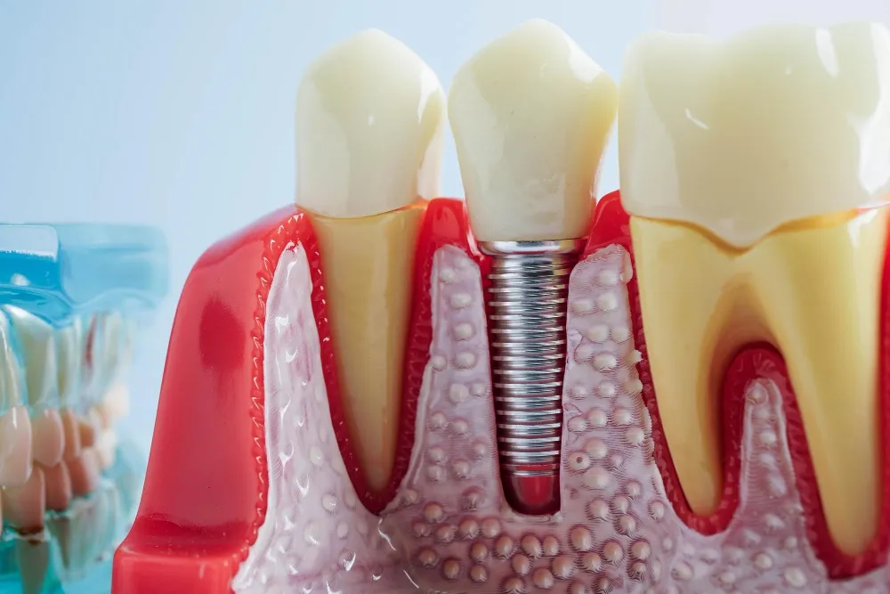
The heart of an implantology procedure is this long-term effort with bone cells of the surface. The mechanical retention (primary stability) achieved by the surgeon tightening the screw into the bone during the surgical procedure is not sufficient on its own for lasting durability. The real strength comes from secondary stability, which occurs over months, namely osseointegration. The titanium materials are specially sandblasted and treated during the production phase. These microscopic pits create an ideal habitat for bone repair cells.
As weeks progress, blood vessels invade the gap between titanium and bone. This system, which carries calcium and phosphorus minerals, builds new spongy bone tissue (woven bone) around the titanium. Within an average period of 2 to 4 months, this spongy tissue calcifies and transforms into a layered, hard, and mechanically resistant (lamellar) bone structure. The only scientific reason for doctors to wait months before proceeding to the prosthesis (crown) fabrication stage is to allow time for this calcification process to complete at the cellular level.
What Methods Should Be Used for Oral Hygiene Practices During the Healing Period?
The oral flora is a moist ecosystem that contains millions of microorganisms by its very structure. When a surgical incision is made, these microorganisms have the potential to penetrate the wound. In the first days following the operation, the area with stitches is extremely sensitive; contact with standard toothbrushes can break the stitches or irritate the soft tissue. Therefore, while the surgical field is exempt from the brushing routine, the remaining teeth in the mouth should be cleaned regularly.
Chlorhexidine gluconate, containing active ingredients, is used for the chemical cleaning of the surgical area in medical mouth rinses. However, there is a critical detail here: the rinse solution should not be vigorously swished in the mouth. Vigorous swishing can dislodge the clot in the wound area. Instead, the liquid should be taken into the mouth and swished gently while tilting the head slightly to allow the liquid to passively wash that area. After the stitches are removed and the epithelial tissue (gum) has closed, normal hygiene routines can be resumed using soft-bristled (surgical) toothbrushes with the physician’s approval.
How Long Does It Take for Stitches to Be Removed and Soft Tissue to Heal?
Epithelial (outer layer) cells are among the fastest renewing cell groups in the body. When the edges of two wounds are brought closer together by stitches, the epithelial cells proliferating from the basal layer migrate towards each other at an average speed of half a millimeter per day, creating a bridge. By the end of the first week, the integrity of the tissue is significantly restored, and the function of the stitches holding the tissue together comes to an end. Since the stitches that remain in the tissue for a long time can pave the way for food and plaque accumulation (infection), doctors gently remove these threads during the clinical appointment they deem appropriate.
Although the removal of the stitches may raise concerns for patients, it is a comfortable procedure that does not require local anesthesia, lasts only seconds, and does not create a sensation in the tissue. Even though the external surface of the soft tissue (gum) heals in two weeks, the tightening and maturation of the connective tissue underneath takes a few more weeks. When the gum reaches its pink, firm, and healthy form, the long-term osseointegration cycle of the underlying jawbone continues safely.
How Does Tobacco Product Use Affect the Biological Repair Process?
In medical literature, smoking is considered one of the most significant environmental factors that affects the success of oral surgical procedures. Nicotine is a powerful vasoconstrictor (blood vessel constrictor). After a surgical operation, the body needs a considerable increase in blood circulation to repair that area. Blood transports the building blocks of cells and the defense mechanisms to the wound site. However, when smoking occurs, blood vessels constrict, the oxygenation of tissues decreases, and carbon monoxide levels increase. The cellular reaction that titanium metal is expected to undergo in poorly perfused bone tissue is delayed.
Moreover, the heat emitted by cigarette smoke creates a drying effect at the suture line, paving the way for the soft tissue edges to open up (dehiscence). The use of tobacco products by an individual in the healing period increases the risk of infection while directly endangering the targeted biological healing. For these medical reasons, limiting smoking a few weeks before the operation and during the cellular healing period in the following months is a parameter emphasized by physicians regarding the safety of the medical process.
How Does Jaw Bone Density Change the Healing Timeline?
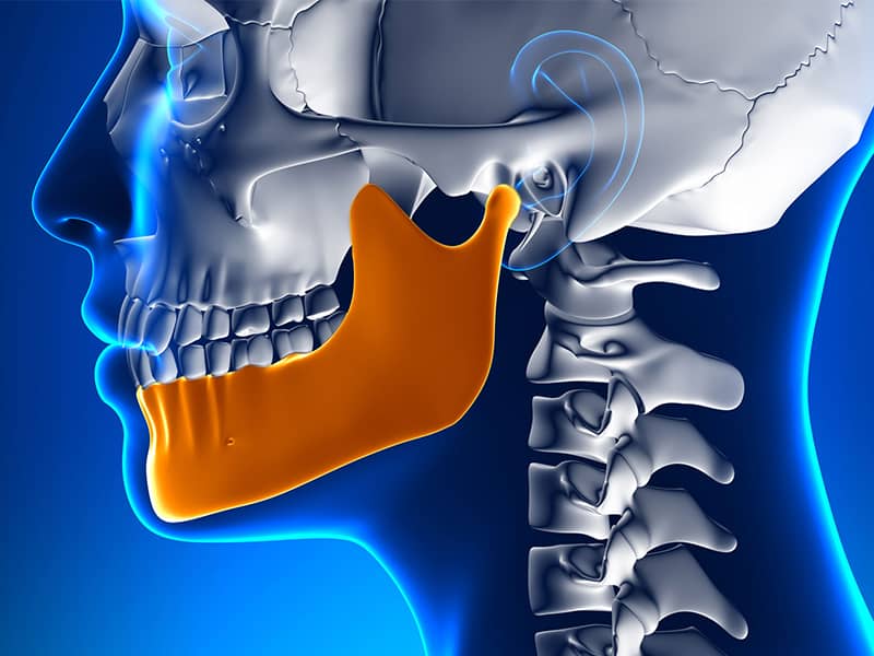
As in different regions of the skeletal system, the densities of the jaw bones are not homogeneous. The hard outer layer of the bone is called cortical bone, while the spongy structure in the inner part is referred to as trabecular bone. The lower jaw (mandible) has a very high calcium density and a dense, hard bone anatomy because it has a moving muscular structure (Type D1-D2 bone). The process of bone-forming cells firmly enveloping the titanium surface within this hard structure takes biologically on average 2 to 3 months.
The upper jaw (maxilla), on the other hand, is a softer, sponge-like, and porous form of bone (Type D3-D4 bone) that is fixed to the skull and contains air chambers such as sinuses. It takes longer for cells to integrate with the bone in this porous area compared to the lower jaw. Physicians often allow for a longer biological maturation period of about 3 to 4 months in upper jaw operations before transitioning to porcelain tooth fabrication, considering this anatomical difference. Additionally, if bone powder (graft) has been used, this timeline extends for both jaws. The table below summarizes this situation:
| Anatomical Area | Bone Density Type | Average Healing (Waiting) Time |
|---|---|---|
| Lower Jaw (Mandible) | Cortical, Dense, and Hard (D1-D2) | 2 – 3 Months |
| Upper Jaw (Maxilla) | Sponge-like and Porous (D3-D4) | 3 – 4 Months |
| Areas with Added Bone Powder | Reconstructed (Grafted) | 4 – 6 Months (Depending on the size of the defect) |
How Do Medical Supports Used After Surgery Affect Healing?
Oral surgery is performed in an area that naturally harbors many bacteria; thus, prophylactic (protective) or therapeutic medications are an important link in the healing chain. The use of antibiotics limits the proliferation of opportunistic bacteria that could penetrate into the bone when the wound is opened, allowing the body’s healing system more time for tissue repair. Strict adherence to the schedule and duration of the prescribed antibiotics is essential to maintain stable medication levels in the bloodstream.
Non-steroidal anti-inflammatory drugs (NSAIDs) not only alleviate pain but also primarily manage the edema and inflammation resulting from surgical trauma at a chemical level. The main reason patients do not experience significant sensitivity post-procedure is that these molecules slow down the release of pain-signaling chemicals like prostaglandins. When medications are administered according to the dosage and duration specified by the physician, they support systemic physiology.
Which Physical Activities Should Be Avoided During the Recovery Process?
Every moment of physical effort exerted by the body shows an increase in heart rate and consequently in blood pressure (tension). In the initial phase of recovery, the vessels in the surgical incision area are protected by a delicate layer of clot. Increased blood pressure directed towards the head region during intense cardio exercises or weight training can push this newly formed clot, leading to seepage (secondary hemorrhages) or a throbbing sensation at the wound site. Therefore, the first 3 to 5 days following the operation should be planned as a resting period away from strenuous activities.
Additionally, environments with high temperatures such as baths, saunas, and steam rooms trigger edema (swelling) by causing vasodilation (expansion of blood vessels). If advanced surgery, such as sinus lifting in the upper jaw, has been performed, activities that cause changes in atmospheric and hydrostatic pressure, like swimming or diving in the sea, are generally restricted for several weeks during the recovery period, as they apply mechanical stress to the sinus membrane. Light-paced walks are among the activities that do not pose a risk since they support circulation.
Healing Abutment (Gingiva Former) Which Stage Is It Attached For?
During the initial stage of the surgical procedure, the titanium root placed inside the bone may be completely covered by gum tissue to protect it from infections. Months later, when osseointegration is confirmed radiologically, the transition to the prosthesis (crown) phase occurs. However, if the crown is placed directly on a flat gum tissue, it anatomically does not sit naturally and is prone to food trapping. To prevent this, a small local intervention is made to millimetrically open the closed gum tissue, and cylindrical metal parts are screwed onto the titanium root.
These parts are referred to as gingiva former (healing abutment). They are generally kept in the mouth for 7 to 14 days. The purpose of this duration is to allow the flat gum tissue to wrap around this metal cylinder, forming a hollow shape resembling a chalice rather than flat. Once the gum takes this shape, the final zirconia or porcelain tooth prepared in the laboratory fits perfectly into this cavity; it creates a biological and aesthetic integrity as if a natural tooth has emerged from the gum in its original form.
How are Temporary Prostheses Used During the Healing Period?
Especially in cases of loss in the front area (smile line) or complete edentulism, the prolonged waiting period without teeth can pose psychological and social challenges for patients. To make this process comfortable, temporary acrylic or clear plate type prostheses are designed that do not transmit pressure to the titanium root (non-functional). In cases of single tooth loss, light-winged devices that attach to adjacent teeth are used to visually fill the gap.
If all the teeth are missing at the hospital and there is a previously used removable partial denture, after the operation, the inside of this denture will be covered with a special soft liner and readjusted to fit into the jaw. The soft liner prevents irritation over the stitches and restricts hard impacts on the bone. The fundamental medical rule for these temporary structures is to only fill the space aesthetically without being included in the functions of hard chewing. To avoid disturbing the delicate connections between the titanium and the bone, patients should refrain from consuming hard foods with temporary teeth.
How is the Recovery Period Follow-Up Planned at Avrupadent Clinics?
Monitoring this biological repair timeline is an integral part of the clinical procedure to manage complications before they arise. At Avrupadent, the first control appointment is usually organized on the 7th or 10th day after the surgical procedure. During this brief appointment, the status of the gum cells’ merging (epithelialization) is observed, any suture threads are removed if present, and evaluations are made regarding the patient’s hygiene routine.
During the waiting months following this, patients are called for radiological follow-ups to determine whether they are ready for the prosthesis stage. The calcium density between the bone and titanium material is analyzed in the X-rays or CT scans taken. If necessary, the stability (ISQ) value of the piece within the bone is tested with a mathematical measurement using special frequency devices such as Resonance Frequency Analysis (RFA). When biomechanical data reaches safe reference ranges, transition to laboratory and measurement stages is ensured in accordance with medical guidelines.
Frequently Asked Questions (FAQs)
1. Is it normal to experience bleeding on the first day after surgery? It is a normal physiological response for mild pink-colored blood to seep from the incision during the first 24 hours following the surgical procedure. As long as the bleeding does not become intense, it does not constitute a medical issue to be concerned about. 2. When does swelling reach its peak and when does it decrease? Swelling due to tissue trauma typically peaks 48 hours post-procedure. After this period, the swelling gradually decreases as the cells distribute their fluids (reabsorb). 3. When can I brush the stitched area normally? Until the soft tissue closure of the surgical area is ensured, it is necessary to protect the area from brushing. Generally, after the stitches are removed and approval is given by the doctor (approximately 2 weeks), the area can be cleaned with extra soft (surgical) brushes. 4. Is there a way to shorten the bone healing process? Osseointegration is a biological process dependent on the metabolic activity rate of osteoblast cells. Although the patient’s vitamin D and calcium balance supports this process, since cellular calcification requires a certain amount of time, it cannot be significantly accelerated from outside the medical timeline. 5. Does air travel hinder the healing process? In standard procedures, air travel does not pose any dangers from the perspective of air pressure. However, if an advanced surgery such as sinus lifting has been performed, changes in atmospheric pressure may negatively affect the healing, thus limiting air travel for medical reasons in the initial weeks. 6. How should the consumption of hot and cold beverages be balanced? During the first three days of healing, excessive hot beverages should be avoided as they can cause vasodilation and trigger bleeding; thus, room temperature (lukewarm) foods should be chosen. Excessively cold foods can create sensitivity, so a thermal balance should be sought. 7. Does the addition of bone powder (graft) create a painful healing period? The addition of a graft does not change the procedure itself, only the volume of the bone. Since the procedure is done under local anesthesia and is managed afterward with medical instructions (medications), it does not create extra pain; it only extends the biological waiting period. 8. Does alcohol consumption disrupt tissue repair? Alcohol can slow down the blood clotting mechanism in the wound area and may lead to dehydration in tissues. Additionally, since it can interact with prescribed antibiotics, consumption should be limited in the first weeks. 9. If the stitches come loose, does it mean the wound has opened? An absorbable suture can typically break down on its own within 7 to 10 days as it is decomposed by enzymes in the body. As the tissue is already healed during this time, it does not mean that the wound has reopened unless the doctor determines otherwise. 10. Does sleeping position affect edema formation? Keeping your head slightly elevated on the night of the operation (for example, using two pillows) is a physical measure that reduces blood pressure in the head area, limiting fluid accumulation and edema formation. 11. How can we understand that the bone integration has been successful? Once the waiting period of several months is over, dental X-rays will show that the bone density has enveloped the titanium surface. Additionally, the stability measurements and light touch (percussion) tests conducted by the doctor during the clinical examination confirm the success. 12. Is it necessary to suture again when the healing cap is placed? Since the procedure of placing the healing cap (gingiva former) is performed with a very localized and millimetric gum margin, it usually does not require sutures and is carried out in a very short clinical session. 13. When does the bruising (ecchymosis) in the surgical area go away? The color changes (bruising) associated with capillary leaks under the skin will gradually fade and disappear on their own within an average of 7 to 10 days, as the fluid is absorbed by the body’s lymphatic system. 14. Is the dislodgement of the blood clot (dry socket) dangerous? In the early days, detaching the clot in the Yara region through mechanical effects such as vigorous rinsing or suction (using a straw) can lead to exposure of the underlying bone (alveolitis); this situation necessitates a medical dressing by the doctor. 15. Should food supplements or vitamins be used during the healing period? In addition to regular nutrition, patients with systemic deficiencies of vitamin D or calcium should take these supplements with the approval of their relevant healthcare providers, as it can provide biological support for bone repair metabolism. |