How is Anterior Dental Aesthetics Achieved?
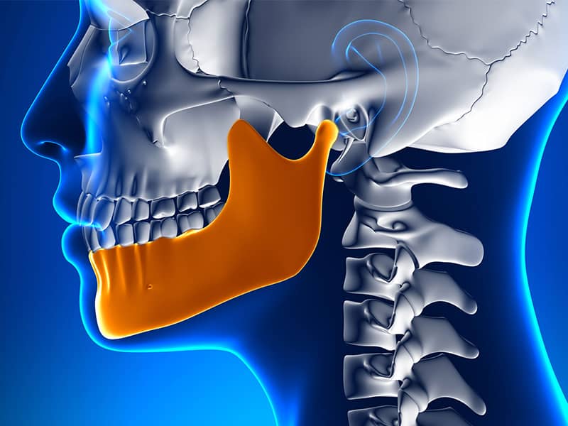
The incisors and canine teeth located in the front part of the mouth, as well as the dental and jaw structure, are among the most strategic anatomical components that directly affect the overall expression of the human face, speech mechanics, and the symmetry of a smile. The teeth in this area, referred to as the “anterior region” in medical literature, are not only responsible for the function of tearing food; they also provide physical support to the muscles of the upper and lower lips, limiting the collapse of the vertical dimension of the face and the formation of an aged appearance. Changes in color, enamel cracks, anatomical asymmetries, or tooth deficiencies occurring in the front area lead to clinical conditions that directly affect individuals’ communication processes in their social lives. The process of restoring such biomechanical and visual losses is structured with multidisciplinary approaches based on current Aesthetic Dentistry principles.
In evaluations made according to the clinical standards of Avrupadent, it has been observed that anterior restorations cannot be limited to mere material selection. The optical properties of the porcelain or composite to be used, the symmetrical curves of the gums (zenith points), and the mathematical ratio that the dental form establishes with the patient’s facial lines are all considered as a whole. These approaches aim to establish a biocompatible, long-term biological balance that is suitable for the individual’s occlusion (jaw closure relationship). In this detailed medical guide we have prepared, the sources of the fundamental issues encountered in the front teeth, the technical properties of porcelain veneer and zirconium applications, the contribution of digital measurement processes to planning, and the scientific criteria under which oral compatibility is achieved are examined.
What Causes Aesthetic Problems in the Anterior Dental Region?
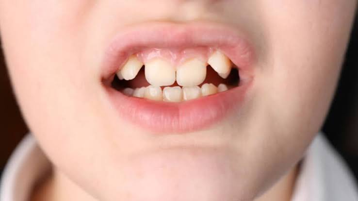
The front teeth are the most prominent anatomical structures inside the mouth and are most exposed to external impacts. Mechanical traumas such as blows, falls, or hard object impacts can lead to tiny cracks or significant tissue ruptures, especially on the incisal edges of the upper central incisors. The dentin tissue exposed beneath the fractured enamel layer gradually absorbs external color molecules over time, resulting in unwanted color discrepancies in the tooth. Additionally, a developmental anomaly known as “peg lateral,” where the lateral incisors are conical and much smaller than normal, is one of the hereditary issues that deeply affects the symmetry of the front region.
Darkening of tooth color is the result of various chemical processes. Anterior teeth that have undergone root canal treatment and have lost their vitality gradually lose their internal fluids over time, becoming dull and taking on a gray/brown tone (non-vital tooth discoloration). Tetracycline antibiotics used in high doses during the developmental stage or excessive fluorides in drinking water (fluorosis) penetrate the crystalline structure of enamel and create deep stains in the tooth’s own tissue in the form of horizontal bands. Additionally, gaps that occur between teeth due to the genetic mismatch of jaw width and tooth dimensions (diastemas) can disrupt airflow, leading to phonetic (speech production) problems.
How is the Proportion of Anterior Teeth Determined in Smile Design?
The anatomical harmony of a smile is dependent on the mathematical balance established between the sizes of the teeth themselves and the facial contours. This balance is shaped by the principle of the “Golden Ratio” (1.618) in aesthetic planning. In the individual’s frontal appearance, the horizontal width of the upper first incisor should optically decrease at a certain ratio as it progresses toward the adjacent second incisor and the canine. The lateral teeth create a sense of depth, appearing positioned further back compared to the central teeth, thus supporting the perspective perception of the face.
When looking at the individual’s singular tooth ratio, it should be noted that the ideal ratio of the width to height of the upper central incisor (central tooth) must approach clinical standards, which are approximately between 75% and 80%. If this ratio is disrupted and the width of the tooth approaches its height, the tooth takes on a “square” and bulky appearance. Additionally, gender and age characteristics are integrated into the process when determining tooth forms. Sharper and straight-edged tooth forms create a masculine expression by emphasizing a strong jaw structure, while rounded (softened) and translucent-edge tooth forms result in a more feminine and youthful facial expression. The digital analyses applied in the Avrupadent process management guide planning phases by translating these facial proportions into mathematical data.
In What Situations Are Porcelain Veneers Preferred for Anterior Teeth?
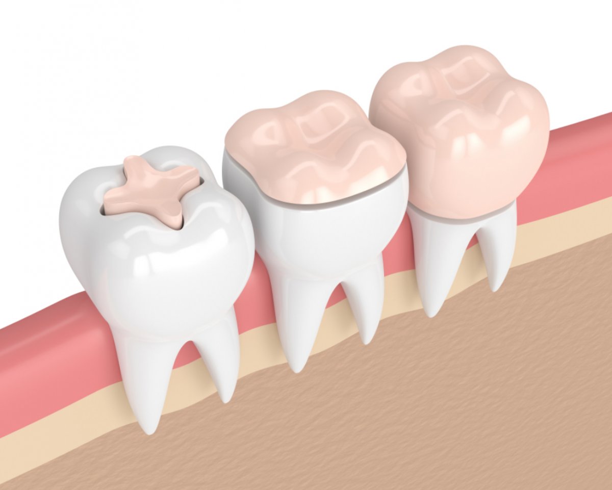
Aesthetic Dentistry is increasingly important, embracing the principle of minimal intervention on tissue integrity (minimal invasive approach) every passing day. Instead of completely cutting around the tooth, porcelain veneers are applied by removing a very thin layer of enamel, approximately 0.3 to 0.7 millimeters, only from the front surface (vestibular area). These leaf-like thin layers, made from lithium disilicate or feldspathic glass ceramics, optically mimic the light transmissibility of natural tooth enamel.
In cases where there is form asymmetry in the front teeth but without significant pathology in the jaw closure relationship (occlusion), porcelain veneer applications yield quite effective clinical results. When these finely prepared ceramic sheets are chemically bonded to the tooth with special medical resin cements (bonding agents), they possess a breakage resistance similar to that of natural tooth enamel. Since their surfaces are factory polished and non-porous, they create a structural barrier against dental staining agents such as tea, coffee, or tobacco.
What Role Do Zirconium Crowns Play in Anterior Area Restorations?
In cases where dental tissue has been lost extensively due to decay or trauma, and thin porcelain laminates cannot adhere or have a risk of breakage under chewing pressure, zirconium crowns that encase the tooth 360 degrees from every angle are included in the planning. Zirconium dioxide is a white and crystalline element that replaces the gray metal substructure embedded inside traditional porcelain crowns. This element ensures biological compatibility by eliminating the dullness caused by metal and the bruising that occurs in the gums (metal reflections).
The latest zirconium blocks produced for anterior region restorations are materials that have maximized light transmittance (translucency) by increasing the cubic phase ratio they contain. When a dark gray or brown root color exists in the tooth’s substructure, zirconium masks this dark color, delivering a bright whiteness to the upper layer. This material, which possesses extremely high biocompatibility, does not provoke a foreign body reaction from the gum tissue; thus, a healthy and stable pink-white tissue integration is established in the collar area where the crown meets the gum.
| Comparison Parameter | Porcelain Laminate (Veneer) | Zirconium Crown |
|---|---|---|
| Intervention on Dental Tissue | Only minimal (0.3-0.7 mm) enamel reduction is done from the front (lip) surface. | The tooth is reduced 360 degrees from every angle (approximately 1-1.5 mm) to create a supportive substructure. |
| Mechanical Strength Structure | It gains strength when chemically bonded, but is fragile when alone, resembling broken glass. | It has a high resistance to external forces and structural stiffness on its own. |
| Clinical Application Area | Color inconsistencies, small diastemas, form adjustments, and aesthetic smile design. | High tooth loss for darker roots, masking of dark-colored roots, and bridge restorations. |
How is the Composite Bonding Method Applied on Front Teeth?
Composite resin applications developed for patients who do not want any loss of tooth tissue but complain about small cracks, gaps, or length discrepancies in their front teeth are fundamental components of the protective aesthetic concept. This method, which does not require a laboratory phase, is completed in the same session as the direct restoration performed by the dentist. After the tooth surface is etched with micro acids, bonding liquids that ensure adherence to the tooth are applied.
Afterward, nano-hybrid composite materials selected from different types according to the natural outer color of the tooth, its transparency, and dentin opacity are added to the tooth in small portions (layering technique). These are polymerized (hardened) with halogen or LED light devices, providing an organic bonding to the tooth. In the final stage of the process, special polishing disks and pastes are used to achieve a glass-like shine on the composite surface. Composite bonds can easily be repaired with a practical material addition in the clinical chair without having to remove the entire restoration in case of any minor chipping or wear that may occur in the future.
How are the Color Changes in Anterior Teeth Managed with Whitening (Bleaching)?
No matter how perfect the alignment and shape of the anterior teeth is, the dark pigmented markings trapped in the dentin tissue beneath the tooth enamel shadow the overall brightness of the tooth. The office-type whitening procedure, which is one of the Aesthetic Dentistry procedures, is a medical chemical process that repairs these color disturbances without making structural cuts. During the procedure, the gums of the patient are protected from the chemical agent using a specially hardened barrier.
The high concentration hydrogen peroxide whitening gel is carefully applied to the front surfaces of the teeth. When activated by laser or high-intensity light sources, it releases free oxygen radicals that penetrate through the microscopic pores (gaps) of the enamel and break down long-chain dark color molecules, converting them into colorless structures. When supported with transparent splints and lower concentration gels (home bleaching) prepared for use in the home environment, the stability of the achieved shade can be maintained for months and years.
How Does Pink Aesthetics Affect the Appearance of Front Teeth?
Gum aesthetics is not just about the shape of the teeth, often referred to as “white aesthetics”; the “pink aesthetics” (gum tissue) that surrounds the teeth ensures that the teeth are perceived as part of a frame. When individuals smile, if the upper lip is raised too much or if the gum covers more of the tooth crown than necessary, it creates a clinical picture known as “Gummy Smile” (gum smile). In this case, the teeth appear quite short, square, and embedded.
To eliminate these asymmetries, periodontal procedures (gingivectomy or gingivoplasty) are employed. Using diode laser technologies or electrocautery, excess gum tissue is cut at a millimetric level and raised to reveal an appropriate crown length (aesthetic) in accordance with the anatomical root lengths of the teeth. The use of lasers during the procedure minimizes bleeding, making wound healing quite comfortable. After the gum margins are equalized according to the golden ratio rules, aesthetic integrity is completed by integrating porcelain laminates or crowns onto this new framework.
What Methods Are Used for Anterior Region Restorations in Tooth Deficiencies?
Loss of anterior teeth due to trauma or periodontal causes leads to a rapid collapse (resorption) in bone volume and gum curvature. In surgical artificial root treatments applied to rectify this deficiency, it is vital clinical parameters to place the titanium fixture into the jawbone at the correct three-dimensional angle. The emergence profile of the zirconium superstructure to be attached to the artificial root should give the impression of emerging just like a natural tooth, which is the most critical aesthetic threshold for anterior region restorations.
In cases where the jaw is not suitable for surgical operation, metal-free (zirconium or full ceramic) bridge systems prepared by taking support from neighboring teeth in the edentulous area are applied. The area where the body (pontic) of the bridge rests on the gums is shaped with ovate pontic (egg-shaped) designs. This design creates an anatomical illusion by lightly pressing the porcelain against the gums as if it is growing out from inside the gums and prevents the loss of gum papillae.
How Do Orthodontic Treatments Prepare for Anterior Aesthetics?
In some cases, the crowding (positioning disorder) of the anterior teeth or gaps between the teeth may be so severe that they cannot be corrected solely with restorative materials (crowns or laminates). Attempting to align a tooth that remains outside the arch using porcelain requires cutting (preparing) much deeper than the enamel layer. This situation may lead to exposing the tooth’s nerve (pulp) chamber, necessitating root canal treatment, and result in the tooth losing its vitality.
In accordance with the protective principles of dentistry, orthodontic specialists integrate the process to avoid such extensive material loss. Teeth are gradually moved into their correct geometric positions by applying short-term clear aligner or bracket treatments, allowing the roots of the teeth to shift slowly within the bone. After the teeth are aligned, an aesthetic specialist performs a superficial polishing at a level of only one-tenth of a millimeter to apply porcelain laminates or composite bonding. Orthodontic support serves as a bridge that fundamentally ensures the medical safety of Aesthetic Dentistry restorations.
How is Anterior Tooth Planning Conducted in Avrupadent Clinics?
In the provision of medical services, meeting aesthetic expectations depends on the flawless analysis of functional health infrastructure and predictable clinical protocols. In Avrupadent process management, each step from the patient’s entry into the clinic to the delivery of the restoration is monitored with a specified series of standards. In the initial phase, the dynamics of the patient’s bite (occlusion), the stretching capacity of the lip muscles, and whether there is active inflammation (swelling) in the gums are clinically analyzed.
All findings obtained are converted into three-dimensional (3D) digital models using intraoral optical cameras. These digital models are matched with the patient’s facial photographs in computer-aided design (CAD) software to draw the virtual architecture of the new teeth. A Mock-Up (trial) fitting done without cutting the patient’s teeth using molds derived from 3D printers ensures that the doctor and patient reach an agreement on the final appearance. When transitioning to the laboratory production phase, corporate procedures are adopted, taking into account the biocompatibility standards of the zirconium or glass ceramic blocks used; maintaining function, aesthetics, and biological integrity over extended periods.
What are the Frequently Asked Questions?
1. What methods are preferred for front tooth fractures?
For small fractures that do not harm the tooth, repair is performed in a single session using composite bonding applications, while larger fractures covering more than half of the tooth are planned with lithium disilicate or zirconium crowns for durability.
2. Do porcelain laminates carry a risk of falling off?
Since they are chemically bonded to the enamel using special medical resin cements (bonding agents), porcelain laminates do not exhibit a risk of falling off as long as they are not exposed to abnormal mechanical trauma such as heavy impacts.
3. What is the healing process like after gum shaping?
Since coagulation (bleeding control) is ensured during gum alignment procedures (gingivectomy) performed with diode laser devices, the healing process is generally completed comfortably within a few days.
4. How is color harmony achieved in front tooth crowns?
The color of the tooth enamel is analyzed using digital or manual color scales based on skin tone and age characteristics, and color harmony is achieved by layering special optical dyes into highly light-transmissive (translucent) ceramic materials.
5. What is the main difference between zirconium and laminate?
While zirconium is a material with high mechanical strength that covers the entire surface of the tooth 360 degrees; laminate is a very thin porcelain that is applied only to the front (lip) surface of the tooth, meeting aesthetic expectations.
6. Does composite bonding change color over time?
Since it contains organic polymer compared to porcelain, it can retain surface stains over the years due to the consumption of strong tea, coffee, and tobacco; however, it can be returned to its original shine with a short polishing process performed in a clinical setting.
7. What restoration is done on a root-treated front tooth?
To mechanically protect the front teeth that turn gray/black after root treatment and to mask the dark color due to fluid loss, zirconium-based full ceramic crowns that surround the tooth completely are planned.
8. Does tooth whitening harm the enamel layer?
Peroxide-containing gels applied under the control of a physician at medical standards do not erode or thin the enamel surface; they only oxidize and break down dark pigments in the dentin layer by filtering through the pores.
9. Can gaps between teeth be closed without orthodontics?
If the general alignment of the teeth is correct, the anatomical gaps between the front teeth (diastema) can be closed proportionally with porcelain laminates or composite resins added without any intervention to the tooth, without the need for any wire treatment.
10. What is the margin of error in digital smile design?
Since computer-assisted CAD/CAM systems and intraoral optical scanners calculate at levels below a millimeter, they minimize human-related (manual) measuring and casting errors at the medical level during the production phase.
11. What should be considered for oral care in anterior area restorations?
In addition to the standard brushing routine, to limit the microscopic plaque accumulation at the junction areas where porcelains meet the gums, interdental brushes, dental floss, and, if necessary, the use of oral rinses should be included in the daily hygiene routine.
12. How does the habit of teeth grinding at night affect anterior tooth restorations?
Involuntary and severe teeth grinding (bruxism) activities during sleep can create excessive mechanical pressure on porcelain structures, potentially leading to cracks; using a custom night guard (splint) is a medical requirement to keep this situation under control.
13. Can changes be requested for the tooth during fittings?
Yes, since the Mock-Up (the physical trial of the virtual design in the mouth) stage has not yet completed laboratory production, requests for revisions regarding the patient’s tooth size, angles, or thickness are taken into account and reflected in the design.
14. Does placing a crown on a single front tooth create incompatibility with the others?
Thanks to modern laboratory techniques, restorations that match the overall aesthetics can be successfully made by examining the translucency of adjacent natural teeth, micro-surface porosities, and color gradients.
15. What function do temporary teeth serve during treatment?
After the preparation (grinding) of the teeth, the fitted temporary teeth protect the exposed tissues from external factors and sensitivity, support the shape of the gum health, and prevent aesthetic loss.