How are Digital Measurement Technologies Used in Aesthetic Dentistry?
The restorative processes in the mouth, teeth, and jaw structure that reconstruct the asymmetries, form defects, and color differences according to medical principles have entirely entered a new era with advancements in material science and optical imaging technologies. In previous years, the measurement stages largely depended on the physician’s manual skills and the technician’s visual perception, which have now transitioned to being controlled by digital systems capable of calculations at the micron level. In Aesthetic Dentistry practices, the process of obtaining measurements is not merely about creating a copy of existing teeth; it is also a critical medical map that relays the structure of the gums, contact points with adjacent teeth, and the relationship of the jaw occlusion in a three-dimensional space to the laboratory.
In traditional methods, the use of silicone-based impression materials filled into metal or plastic spoons occupies a volumetric space in the patient’s mouth, and often leads to microscopic deformations due to the intrusion of liquids like saliva or blood during the impression-taking process. In current clinical disciplines, intraoral (in-mouth) optical scanners have replaced these physical materials. This process, which involves the instantaneous conversion of thousands of photographs taken from dental surfaces into a three-dimensional model via computer software, minimizes the margin of error. Transforming the anatomical data of the patient into a digital format allows for the production of planned restorations without manual intervention using CAD/CAM systems and enables virtual design according to facial proportions. In this medical guide we have prepared, the operation of digital impression technologies in a clinical setting, the differences they provide compared to traditional impression methods, and the integration steps in Avrupadent processes are examined in detail.
How Do Intraoral (In-Mouth) Scanners Work in a Clinical Setting?
The working principle of intraoral scanners is based on sending laser or structured light beams to the dental surface and the reflection of these lights returning to the sensors after bouncing off the tissues. The device’s tip is slowly moved by the dentist over the patient’s teeth from a certain distance. By calculating the time taken for the light to hit the surface and return, all indentations, protrusions, chewing surfaces, and junctions of the gums (collecting areas) are recorded along with depth perception. The powerful processors inside the scanner stitch each frame together to create a colorful and three-dimensional model of the mouth within seconds.
In clinical practice, the use of these systems provides immediate feedback to the dentist during the measurement process. For instance, if an area is missed during scanning on the back surfaces of molars or in narrow regions, the software marks this area in a different color on the screen, alerting the dentist. Instead of starting the measurement from the beginning, the dentist can simply move the camera over that missed area again to complete the missing data. This feature ensures the complete recording of the foundational data essential for treatment, increasing the reliability of subsequent design phases.
What Medical Differences Does the Digital Measurement Method Offer Compared to Classic Silicone Measurements?
In traditional dentistry, alginate, polyether, or silicone-based impression materials are obtained by mixing powder-liquid or two different chemicals. When these materials are placed in the patient’s mouth, they undergo a hardening reaction (polymerization). During the reaction, microscopic bubbles may occur depending on temperature changes and humidity factors. Additionally, when the impression is removed from the oral cavity, tears or deformations in the impression may happen due to negative pressure areas, which are common occurrences. In digital optical impressions, since a physical substance is not used, all risks of dimensional changes arising from the physical nature of the material are completely eliminated.
There are also significant differences between the two methods in terms of communication and data transfer. For physical impressions to reach the laboratory environment, courier or shipping processes are required, and once the impression reaches the lab, it is expected that the model will be obtained by pouring inside. On the other hand, digital data (usually files in STL, PLY, or OBJ format) is transferred in seconds to the screen of a technician on the other side of the world as soon as the scanning process is completed through the internet or cloud systems. This eliminates the waiting time for the physical model, allowing laboratory processes to start immediately.
| Comparison Criterion | Classic Silicone Impressions | Digital (Optical) Impressions |
|---|---|---|
| Dimensional Stability | Microscopic volume losses may occur in the impression depending on heat and time. | Since the data is numerical, it does not experience any volumetric changes or tears. |
| Patient Comfort | The contact of the impression trays with the palate may cause discomfort. | It is comfortable and offers a pleasant experience because it is scanned with only a thin camera tip. |
| Archiving | The acquired impressions occupy a wide area on physical shelves. | Files are stored on cloud servers without degradation for a lifetime. |
How are Digital Data Integrated into Aesthetic Design?
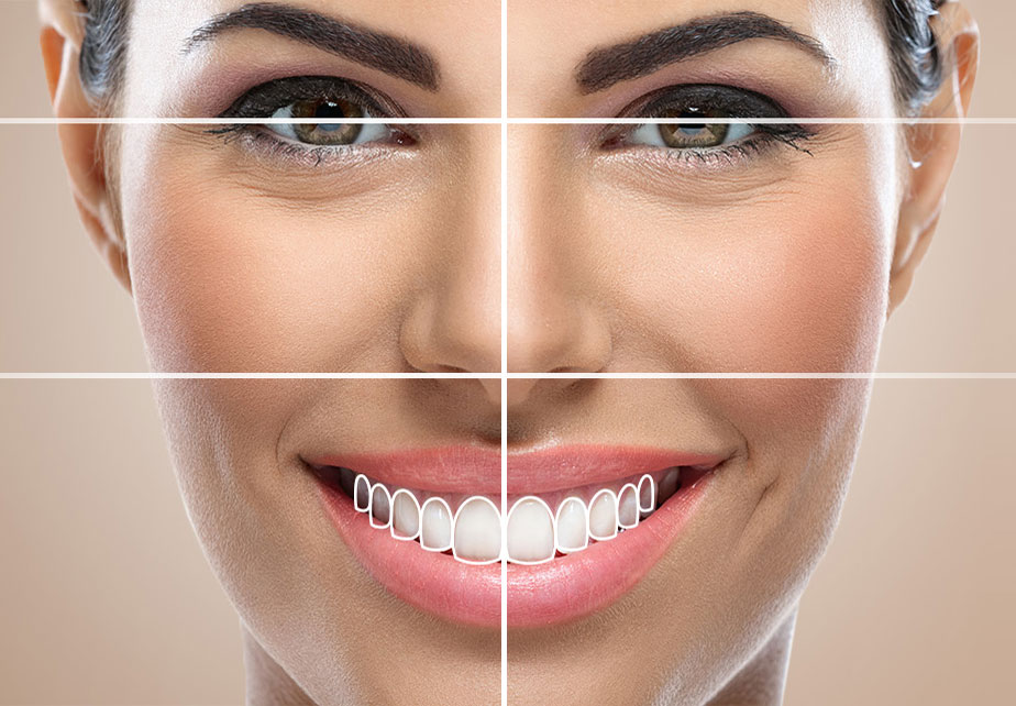
No matter how symmetrical the individual form of the teeth inside the mouth is, aesthetic success cannot be mentioned if they are not compatible with the lips, facial contours, and the mechanics of speech. The true power of digital data in Aesthetic Dentistry procedures comes from allowing this macro-aesthetic analysis. After reflecting the optical measurements onto the computer screen, the physician transfers the photographs showing the resting, smiling, and speaking states of the patient to the same software. The system creates a single digital platform by aligning the intraoral model with the facial photograph based on lip borders.
On this platform, horizontal and vertical lines are drawn referencing anatomical points such as the patient’s pupils (pupillary line), nasal wings, and chin tip. The virtual tooth contours of the new porcelain teeth are positioned according to these lines. For example, in a patient whose lips do not excessively stretch, the length of the front teeth is designed to be somewhat longer to enhance their visibility. All these modifications can be simulated with the help of a mouse in seconds, allowing for a projected design of how the patient’s facial expression will appear at the end of the treatment.
What is the Function of Optical Scans in CAD/CAM Systems?
In the Computer-Aided Design (CAD) phase, the digital impression map sent by the optical scanner becomes the technician’s working surface. The outline of the prepared (reduced) tooth is automatically detected by the software or marked by the technician in the scanning data. The software analyzes the anatomy of the adjacent teeth and the occlusal shape of the opposing arch, offering an ideal tooth form from its library that fits the space. The designer prepares the tooth for function by adjusting the occlusal (chewing) contacts based on this suggestion.
Once the design is approved, the data is directed to Computer-Aided Manufacturing (CAM) machines. These machines are medical versions adapted from industrial CNC devices. Inside the device, factory-produced blocks of lithium disilicate or zirconium, which do not contain air bubbles and are produced as a monoblock, are placed. Robotic micro-mills carve this hard block according to commands from the CAD software, transforming the design into a physical object using a subtractive manufacturing technique. The precision of the optical scanning data is a direct determinant of how successful this milling process will be; as erroneous scanning data cannot produce accurate manufacturing.
How Do Digital Measurements Guide Orthodontic Alignment Needs?
Before starting aesthetic restoration processes, planning an orthodontic preparation phase is a protective medical principle in cases where the misalignment or positional deviation of teeth is significant. Excessive enamel reduction, to cover misaligned teeth with porcelain laminates, carries the risk of damaging the living nerve tissue of the tooth (pulp). In such cases, transparent tray (aligner) treatments come into play.
In transparent tray treatments, digital measurements taken with optical scanners are used to create a movement map of the teeth over weeks. The software simulates how many degrees each tooth will rotate around its axis or how many millimeters it will move, planning the series of transparent trays based on this digital map. Once the teeth reach the desired symmetrical alignment, the dentist applies porcelain restorations by making a very superficial reduction of one-tenth of a millimeter. Digital measurement serves as a common bridge facilitating data exchange between these two disciplines (orthodontics and prosthetic treatment).
What is the Role of Optical Measurements in the Edge Fit of Porcelain Restorations?
The lifespan of a porcelain crown or laminate inside the mouth is determined not only by its resistance to chewing pressure, but also by the tightness of the area where it contacts the gum tissue. The margin line prepared by the dentist on the tooth is the most critical area where the porcelain will adhere to the tooth and come into contact with the gum. If this margin cannot be accurately replicated during the measurement stage, the produced porcelain may either not fit properly on the tooth, leaving a gap (open margin) or press against the gum tissue (overlapping margin).
Fluids and food debris leaking from open margin areas can lead to the natural tooth underneath the crown decaying over time (secondary decay); while overlapping margins continuously irritate the gum tissue, causing periodontal problems such as redness, bleeding, and bone loss. High-resolution lasers or optical readers from intraoral scanners reveal these fine margin boundaries as a distinct line on a digital screen. The design software locks the edges of the porcelain at a micron level to this line. When the edge fit of the produced restoration is perfect, the gum tissue achieves a healthy bond with the porcelain, minimizing biological responses.
Why is Digital Measurement Recommended for Individuals with Nausea Reflex?
One of the procedures patients often hesitate to undergo in dental clinics is waiting for a certain period with a mouth full of impression material. The soft consistency of alginate or silicone-based substances can spread to the back part of the tongue (oropharyngeal region) or towards the soft palate during placement. Stimulating the tissues in this area triggers the central nervous system, activating the body’s defense mechanism known as the gag reflex. This process, which is a stressful experience for the patient, can often lead to the early removal of the impression and a decrease in its quality.
The main parts of intraoral scanners are designed to take up minimal space in the oral cavity anatomically. Since the camera tip only moves over the surfaces of the teeth, there is no mechanical contact with the throat area or the deeper parts of the palate. Additionally, during scanning, the patient can swallow, rest, or the physician can pause the procedure within seconds, allowing the patient to relax. The ability to pause the procedure at any desired moment and continue from where it left off is a medical convenience that makes digital impressions the first choice for patients who experience the gag reflex.
What Physical Forms Do 3D Printer Systems Transform Digital Impressions Into?
In digital workflows, 3D printers hold a significant share among technologies that convert digital designs into physical materials, beyond just computer screens. Unlike CAM devices that work with subtractive methods (cutting), additive manufacturing printers create objects layer by layer by solidifying light-sensitive liquid resins. These devices are used in cases where a physician needs to conduct a trial in the patient’s mouth during the planning of Aesthetic Dentistry.
When the smile design model created in a virtual environment is sent to the 3D printer, it is printed as a jaw model made of resin, identical to the new design. The physician creates a silicone mold over this physical model and performs a “mock-up” trial in the patient’s mouth. Furthermore, to ensure that the patient prepared for restoration does not remain without teeth during the laboratory process and for the purpose of shaping the gums, temporary acrylic teeth can also be obtained with millimetric precision from the same printing systems. Printers serve as a bridge that facilitates the medical team’s work in the materialization of digital data.
How is Jaw Occlusion Analysis Performed in Digital Systems?
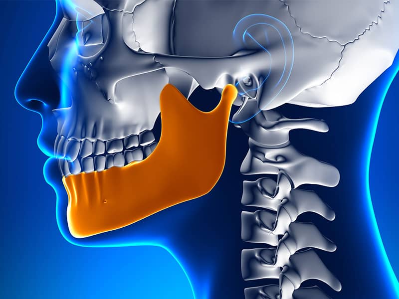
The relationship of teeth contacting each other (occlusion) is one of the building blocks that determine long-term success in prosthetic dentistry. In traditional methods, this relationship is detected using wax or special heating registration materials placed in the mouth; in digital scanners, when the patient closes their jaw naturally, the camera scans the area where both jaws meet from the side. The software accurately positions the upper jaw data with the lower jaw data in the correct occlusal position within seconds based on this side scan.
This virtual locking process provides colorful pressure maps that show how forcefully the teeth touch each other. If there is an area that makes excessive contact with the new porcelain veneer or crown to be made, this area is marked in red. The technician uses a computer mouse to digitally grind this red area to achieve a green (ideal contact) color. Adjusting the closing forces with such care medically prevents porcelain restorations from breaking or causing discomfort in the temporomandibular joint (TMJ) during use.
What Protocols Are Followed in Optical Scanning Processes at Avrupadent Clinics?
It is essential that not only is the technological infrastructure present in clinics, but that these technologies are also applied within the correct medical protocols. In Avrupadent process management, the initial procedure for a patient seeking aesthetic restoration involves cleaning tartar and checking for plaque in the patient’s mouth. If there is inflammation, bleeding, or tissue swelling in the gums, the scanning process is postponed until the gums are healthy and free of inflammation, as it is known that these conditions will change the limits that the optical scanner will detect.
When the documents are properly isolated from the oral environment during scanning, spilling onto the camera lens or the external dental surface can cause topographic errors in the digital data due to the bubbles in the thick fluid. The detailed scanning and facial photograph data obtained are transferred to the clinic’s secure cloud systems. Based on this data, a consultation team consisting of a periodontist, prosthodontist, and aesthetic physician jointly evaluates the planning. The results shared with the patient are harmonized according to expectations and scientific feasibility and proceed to production.
What are the Frequently Asked Questions?
1. Is there a radiation risk when taking digital measurements?
Intraoral optical scanners do not use X-rays; they collect data through high-speed optical photography using only laser or advanced LED light sources, thus they do not expose the body to radiation.
2. Can digital scanning be done during pregnancy?
Since there is no risk of swallowing any chemical measurement material and it does not contain radiation, it can be safely applied during pregnancy when there is no warning from the obstetrician.
3. Does the measurement process take a long time?
Scanning a single arch generally takes between 1 to 3 minutes, depending on the patient’s mouth opening and the area to be treated, making it a quite comfortable and brief clinical procedure.
4. Is digital design suitable for every patient?
Digital planning is suitable for any adult patient whose mouth opening allows the camera to enter and whose systemic health is stable, particularly in the processes of porcelain fillings, crowns, or aesthetic gingival surgeries.
5. How long can digital data be stored in the archive?
The scanning files belonging to the patient (in STL/PLY formats) are preserved on cloud-based servers for a lifetime without any degradation, serving as a resource for potential future reproductions.
6. Will the result on the computer match exactly with the design device?
Thanks to CAD/CAM integration, since the three-dimensional data prepared in the software is produced directly by robotic milling, the dimensions of the designed final prosthesis remain identical at the micron level.
7. Is pain felt inside the mouth during the measurement process?
Since the optical camera scans the surface of the teeth only above a certain millimeter threshold, there is no piercing or cutting contact with the gum or tooth; the process does not involve physical pain.
8. Does swallowing disrupt the scanning process?
The most significant advantage of digital scanners is the ability to pause the procedure whenever desired; when the patient wants to swallow or rest, the camera is withdrawn, and the scanning resumes from the last point with zero error.
9. Do the edges of porcelain restorations exert pressure on my gums?
Thanks to the high resolution provided by the scanners, the junction between the tooth and gum is clearly visible on the computer, and the edges of the produced restoration are aligned at a level that will not exert pressure on the tissue.
10. Can permanent porcelain teeth be printed with 3D printers?
In today’s clinics, 3D printers are mostly used for printing working models or temporary prostheses from resin, while permanent ceramic restorations are produced by milling from blocks in CAM devices.
11. Do digital scanning bite joints (TME) benefit those with discomfort?
Yes, the digital record obtained by closing the hospital’s lower and upper jaws allows for the porcelain to be designed in a balanced way so that they do not apply pressure on the joint, as it shows the occlusal forces on software.
12. Does the treatment duration shorten with digital workflow?
As the steps of sending physical measurements by cargo to the laboratory and waiting for the model are eliminated, the total clinical treatment time significantly decreases since the data reaches the technician within seconds.
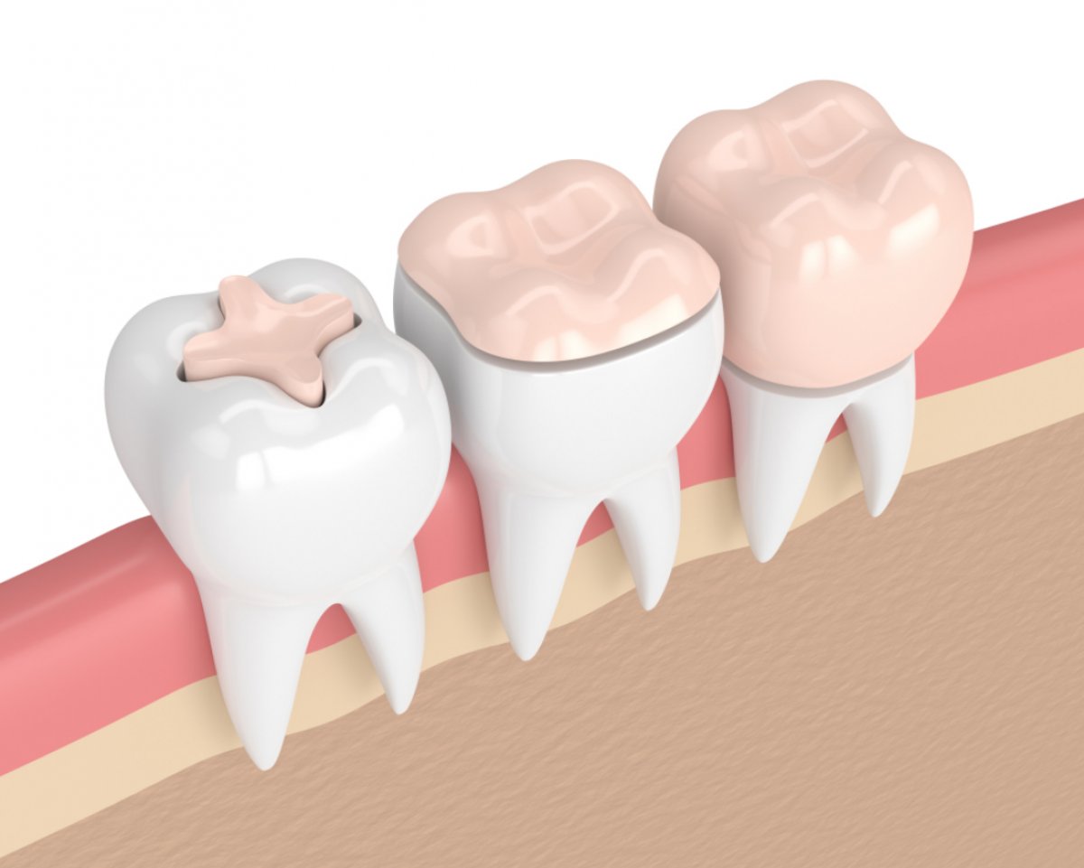
13. Is scanning also used for aesthetic fillings (inlays/onlays)?
In major fracture or break cases, after preparation is completed on the tooth, the measurements taken by the scanner can be used in the laboratory to design and fabricate porcelain fillings (inlays/onlays) which are then applied to the tooth.
14. How is the thickness of porcelain laminates (leaf teeth) adjusted?
The thickness of the laminates designed on the software is determined at an ideal value between the material’s fracture resistance and aesthetic thinness, supported by the computer according to the tooth’s external form and occlusion distance.
15. Are the clear plates used instead of braces also planned with this technology?
Yes, all phases of treatments using clear plates (aligners) to correct misalignments in teeth are planned through virtual movements based on the digital data obtained from intraoral optical scanners.