

How is the implant treatment for patients coming from outside Izmir and abroad progressing?
Compensating for tissue losses occurring in the anatomy of the mouth requires a comprehensive medical logistics planning that encompasses not only physiological limits but also geographical distances. Implant dental applications, based on the principle of placing biocompatible titanium materials into the jawbone, are spread over a specific timeline due to the cellular healing (osseointegration) phase. For individuals traveling to Izmir for health purposes from outside the city or abroad, this timeline necessitates the combination of medical requirements with international travel parameters.
How does the planning of implant dental treatment start for patients coming from outside the city or abroad?
The remote planning process begins with the transmission of radiological images (panoramic X-rays or CBCT scans) that the patient has obtained in their own location, along with their detailed systemic health history, through digital communication channels to the clinic. By examining this medical data, a preliminary medical map suitable for the patient’s anatomical structure is created and the clinical travel schedule is shaped.
In the practice of dentistry, the diagnostic phase, where surgical procedures are analyzed at both microscopic and macroscopic levels, forms the backbone of the operation area. In cases where the patient is not physically present in Izmir, the first step of the diagnostic phase is carried out through digital medical technologies. The patient is asked to go to a imaging center in their city or country to obtain an updated jaw X-ray. The radiographic data transmitted in digital format allows the physician to make a preliminary assessment regarding the density of the bone, the position of the nerve channels, and the angle of neighboring tooth roots.
In addition to radiological data, the patient’s medical history (anamnesis) is collected through standardized digital forms. Medications used, existing chronic diseases, and allergy statuses are recorded at this stage. Based on the obtained anatomical and systemic data, the physician determines the number of titanium screws planned for application, whether there is a need for bone powder (graft), and the medical timeline of the procedure. The coordination teams within İzmir Avrupadent communicate this medical plan to the patient, enabling them to integrate this physiological process into their work or daily routines. The planning is a medical draft prepared to be confirmed with a face-to-face examination to be conducted at the clinic.
What Radiological Examinations and Medical Data Are Required Before Reaching İzmir?
For clinical evaluation, DICOM format three-dimensional Dental Volumetric Tomography (CBCT) or two-dimensional panoramic x-ray data is requested from the patient. Additionally, a list of anticoagulants (blood thinners) used by the hospital, as well as systemic medical reports including diabetes or cardiovascular history, are requested.
In jaw surgery, the thickness of the bone matrix beneath the mucosa and the location of air cavities can only be visualized with radiological devices. In remote data analysis, two-dimensional panoramic x-rays are functional for identifying the general dental arch form and potential cystic pathologies. However, to accurately measure the anterior-posterior (bucco-lingual) width of the jawbone and limit millimetric error margins, three-dimensional tomography (CBCT) data provides significantly higher medical value. For patients coming from abroad, this data is generally requested in the form of files with the international medical imaging standard DICOM (.dcm) extension.
Alongside radiological analyses, the hospital’s systemic laboratory results are also requested. For example, the HbA1c test result reflecting the average blood sugar levels for the last three months is expected from a patient with a history of diabetes. In patients undergoing treatment for osteoporosis (bone loss), information regarding the bone density measurement (DEXA) and prescribed bisphosphonate medications is mandatory. All these medical data are compiled to perform a preliminary analysis of potential physiological reactions that may develop during the surgical procedure and the cellular healing capacity of the bone tissue.
How is the Systemic Health History of Patients from Abroad Evaluated?
The systemic diseases of international patients and the pharmacological agents they use are examined according to universal clinical protocols of the relevant medical branches (cardiology, endocrinology). If necessary, consultations are conducted with the patient’s physician in their home country or with specialists in Izmir to adjust medication dosages suitable for surgical interventions.
While implant surgery is a local procedure, it operates integratively across the entire organism through the circulatory system and the vascular mechanism. International patients often have medical files monitored in different healthcare systems. If the patient is using an anticoagulant (antiplatelet or anticoagulant), there is an increased risk of prolonged bleeding during the incision (cut) on the gum and bone. To manage this physiological situation, approval for a “bridging” (temporary medication adjustment) protocol is sought from the cardiologist in the patient’s home country or from a local internal medicine specialist upon arrival in Izmir.
Similarly, endocrine system disorders (such as uncontrolled diabetes) can slow down osteoblast (bone cell) activity due to impaired cellular oxygenation and blood flow to the wound site (vascularization). The surgical schedule may be suspended until the medical parameters of patients with these systemic findings are regulated to reference ranges. According to Avrupadent clinical procedures, these medical history forms collected remotely will be confirmed with routine measurements such as blood pressure and fasting blood sugar on the first day the patient physically steps into the clinic, concluding the medical preparation phase.
What Anatomical Analyses Are Performed During the First Clinical Examination in İzmir?
During the physical examination in the clinic, the gingival (mucosal) biotype, jaw closure (occlusion) relationships, and the capacity of the oral cavity are measured. The radiological data sent is integrated with the clinical examination to delineate the actual anatomical boundaries of the bone walls and soft tissue thicknesses for surgical planning.
Once the patient completes their travel and reaches the clinic in İzmir, the phase of overlapping the anatomical realities with the virtual planning starts. The bone volume data displayed on the computer screen is considered together with the physiology of the intraoral tissues. The doctor examines the thickness of the gingiva (keratinized mucosal band) in the area where the implant will be placed using periodontal millimetric probes. In patients with a thin (transparent) mucosal biotype, soft tissue grafting may be included in the treatment plan to limit the gray reflection of titanium and prevent bacterial passage.
However, the dynamics of the hospital’s muscles and the movements of the jaw joint (temporomandibular joint) are observed. In international patients, acute muscle spasms (trismus) that may develop due to travel stress or climate changes can restrict mouth opening. By calculating chewing habits, teeth clenching (bruxism) index, and vertical occlusal distance (interocclusal space), it is clarified at which angular vector the dental implants will be placed into the bone. This integrated analysis is a fundamental medical standard that determines the biomechanical compatibility of the operation.
How Many Stages Does Implant Surgery Take for Patients Coming from Abroad?
The medical schedule is generally planned in two different travel cycles. The first visit involves the surgical intervention and the placement of a titanium screw into the bone (averaging 2-3 days); the second visit, which takes place months after cellular integration, includes measurements and the placement of porcelain teeth (averaging 7-10 days).
Dental implant procedures are a biological process expecting the integration of inorganic material with living tissue at a cellular level. Therefore, the procedures consist of stages extending over time, as required by nature. For patients coming from abroad or different countries, these stages are divided into two major periods, during which travel logistics are intertwined with clinical appointments.
In the first travel period, clinical evaluation and surgical operation are performed. Osteotomy (bone drilling) under local anesthesia and the placement of the screw take one or several hours. In the first couple of days following surgery, the patient’s wound healing and possible physiological swelling are monitored by the physician at the clinic. If no complications arise, the patient can return to the city or country they live in. After this stage, the “osseointegration” (the titanium being enveloped by bone cells) phase begins, which lasts for months depending on the anatomical structure. Once cellular maturation is achieved, the patient comes to Izmir for the second time. In this final visit, the cover screws on the gum are opened without any surgical procedure, measurements are taken, and the crowns/bridge prostheses produced in the laboratory are adapted into the mouth, finalizing the treatment at a functional level.
| Visit Phase | Medical and Surgical Procedures Performed | Recommended Duration of Stay in the City |
|---|---|---|
| 1st Visit (Surgical Stage) | Clinical examination, local anesthesia, placement of the implant in the jaw, control of swelling and bleeding in the first 48 hours. | Average 2 – 4 Days |
| Biological Waiting Period | The cellular healing period the patient experiences in their own country/city. | Lower jaw: 2-3 Months | Upper jaw: 3-6 Months |
| 2nd Visit (Prosthetic Stage) | Attachment of healing abutments, occlusal measurements, laboratory trials, and delivery of crowns/prostheses. | Average 7 – 10 Days |
How is the Waiting Period for Integration of the Implant into the Jawbone (Osseointegration) Monitored from Afar?
During the biological repair months that the hospital spent in its own country or city, communication continues through digital systems. While the soft tissue healing of the operative area is monitored through photographic data, the course of cellular regeneration is medically analyzed with periodic X-rays taken and sent to the clinic from the hospital’s location.
The osseointegration phase that begins after the surgical placement procedure is a bone formation process characterized by the organization of osteoblast cells around the titanium threads, accumulating calcium and phosphate minerals. When the patient leaves İzmir and returns to their own geography, this physiological clock continues to work. The closure of the wound site (mucosal incisions) occurs within the first few weeks (epithelization), while cellular stabilization within the bone takes months.
In the remote monitoring mechanism, the patient digitally transmits the superficial visual status of the surgical area to the clinic for the first 15 days. In cases where stitches need to be removed, the patient may consult a local dentist in their own area (if their own dissolvable stitches have not been used). Just before planning the second visit (e.g., 3 months after the operation), the patient is asked to undergo a current radiographic imaging (panoramic X-ray) in their own area and send it digitally to the İzmir Avrupadent clinic. The doctor examines the radiolucency (black area) indicating the situation between the titanium screw and the bone on the X-ray and identifies the radiological findings of the bonding phase and gives medical approval for the second visit.
How Do Bone Powder (Graft) Procedures Affect the Duration in Patients Coming from Abroad with Jawbone Insufficiency?
Bone augmentation procedures (graft/bone powder addition) performed in cases where the alveolar bone volume is insufficient in vertical or horizontal alignment add additional months to the existing biological healing timeline for anatomical repair. Treatment planning may extend over three separate travel cycles depending on the size of the defect.
Years without teeth or cystic pathologies lead to cellular resorption in the jawbone, eliminating the sufficient bone wall to support the implant. To restore these anatomical narrownesses, xenograft (animal-derived) or alloplastic (synthetic) graft particles are placed in the surgical area. In areas of the upper jaw where there are lower teeth, the maxillary sinus membrane that hangs down is pushed up (sinus lifting) and bone powder is applied to the gap.
In minor (small) bone deficiencies, the grafting procedure can be performed on the day the implant is placed; in this case, the travel schedule maintains its standard course. However, in major (large) bone defects, the titanium screw cannot provide initial anchorage (primary stability) in the bone. In the face of this anatomical handicap, treatment is divided into three travel stages: The first visit involves only the bone powder (augmentation) operation, and an average of 4 to 6 months is waited for the graft to integrate with the patient’s own viable bone (angiogenesis and mineralization). In the second visit, the implant is placed in the newly formed bone (it is expected to integrate as well). In the third visit, prosthetic (crown) teeth are installed. This medical requirement is a cellular reality that radically affects the logistical planning of patients coming from abroad.
How Many Days Does the Prosthetic (Crown) Stage Affect Travel Planning?
The prosthetic phase that begins once the periosteal integration is complete requires an average clinical schedule of 7 to 10 days, including gingival shaping, occlusal measurement acquisition, laboratory production, and mechanical trials. The duration of patients’ stay in Izmir is determined according to the scope of the restoration (single tooth or entire arch).
The second trip to Izmir does not involve a surgical osteotomy (bone drilling) procedure but encompasses a meticulous engineering and laboratory process. The gingiva covering the screws that are integrated with the jawbone is locally opened, and the healing caps that will shape the mucosa emergence profile are placed. The mucosal tissue takes on its physiological shape around this cylindrical metal within a few days. Once the gingiva matures, a three-dimensional copy of the jaw curves is transferred to the dental laboratory using digital intraoral scanners or conventional measuring materials.
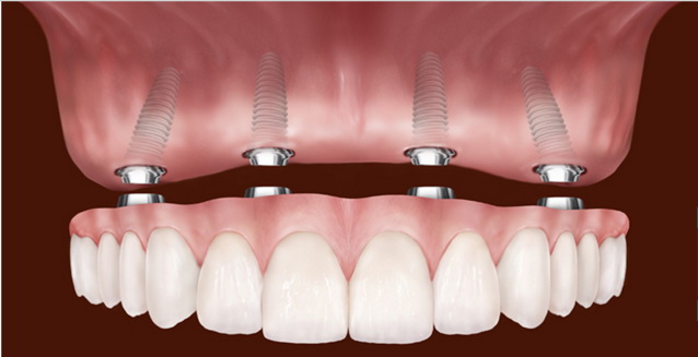
The laboratory phase includes the milling and baking procedures of zirconium or metal-supported ceramic crowns that will optimally transfer the masticatory forces (occlusion) to the axis of the screw. The mechanical compatibility of the ceramic teeth with the hospital’s oral flora, lip support, and contact points with opposing teeth is adjusted step by step through clinical trials. In restorations involving one or several teeth, this process takes approximately 7 days; in complex fixed restorations such as ‘All-on-4’ covering the entire arch, establishing masticatory balance may extend up to 10 days. Patients are required to organize their return tickets considering the usual durations of this medical production phase.
Does Air Travel Affect Biological Tissues After Implant Surgery?
Standard implant surgeries typically do not adversely affect cellular tissues during air travel. However, in upper jaw surgeries where the “Sinus Lifting” (sinus membrane elevation) procedure is applied, changes in cabin pressure (barotrauma) can cause damage to the membrane, so a certain physiological waiting period should be observed before flying, with the doctor’s approval.
One of the frequently encountered medical conditions in travel logistics is the physical effect of cabin pressure on the surgical wound site. In standard artificial root surgeries placed in the lower or upper jaw, the surgical procedure applied to the bone is a closed wound system (the gum is sutured closed). Altitude-related pressure differences experienced during commercial flights do not lead to acute physiological complications in this closed bone matrix. In related cases, patients can join their normal flights 24 to 48 hours after the surgery (when clot stability is ensured).
In contrast, the medical approach varies when the “Sinus Lifting” procedure is applied in the posterior (molar) region of the upper jaw. The maxillary sinuses are anatomically connected air-filled cavities associated with the nasal cavity. When the membrane of this organ is surgically lifted and grafted, the region becomes extremely sensitive to pressure changes (Boyle-Mariotte law). Changes in air pressure during flight can cause the gases within the sinus to expand (barotrauma), potentially leading to ruptures in the newly repaired sinus membrane or severe localized pain. For this biomechanical reason, it is a medical requirement for patients who have undergone sinus surgery to remain at sea level for about 7 to 14 days before flying or to have flight plans approved by their physician.
What Medical Rules Should Be Followed During the Recovery Period After Returning from Abroad?
When patients return home, they must use the antibiotics and chlorhexidine mouthwashes prescribed by the physician for infection prophylaxis according to their medical schedules. It is essential to avoid smoking, which suppresses tissue circulation and hinders bone synthesis, for cellular healing.
In the first days after the operation, mild swelling and capillary leakages that appear are part of the body’s inflammatory repair response. When international patients return to their regions, they should temporarily redirect their chewing reflexes to the jaw area that hasn’t undergone any procedures to protect the anatomical integrity of the wound site (mucosal incisions). Extremely hot and acidic beverages can cause leaks in the surgical area by dissolving the clot structure (fibrin network), so a puree diet model should be adopted at room temperature.
The major factor negatively affecting wound site physiology is tobacco products. Carbon monoxide in cigarette smoke binds to hemoglobin with a higher affinity than oxygen, reducing tissue oxygenation (hypoxia). Simultaneously, nicotine constricts peripheral (capillary) vessels, limiting the amount of blood reaching the surgical site. A wound that does not receive sufficient nutrients and defense cells has difficulty closing and becomes open to bacterial growth. In light of these physiological facts, controlling tobacco consumption during the osseointegration process is a clinical rule.
How is Long-Term Implant Care and Clinical Management of International Patients Carried Out?The functional lifespan of implant-supported restorations is dependent on mechanical oral hygiene provided using special dental threads and auxiliary brushes, in addition to standard brushing. International patients should consult local dentists in their countries every six months for professional tartar (calculus) cleaning and to measure the radiological bone levels.
The functional lifespan of implant-supported restorations is dependent on mechanical oral hygiene provided using special dental threads and auxiliary brushes, in addition to standard brushing. International patients should consult local dentists in their countries every six months for professional tartar (calculus) cleaning and to measure the radiological bone levels.
Titanium material does not crack under acid attacks, similar to natural tooth enamel containing calcium hydroxyapatite crystals. This inorganic structure may create a false perception that there is no risk of infection. Conversely, the gum tissue (peri-implant mucosa) and alveolar bone surrounding the titanium screw are much more fragile against bacterial dental plaque compared to natural teeth. The periodontal fiber networks present in natural teeth that slow infections are not present around implants (ankylosis structure).
Due to this anatomical difference, if the bacterial plaque accumulating around the porcelain tooth (at the margin line) is not cleaned, soft tissue inflammation (peri-mucositis) can quickly progress to the jawbone, leading to bone resorption (peri-implantitis). Patients who complete their treatment and leave İzmir are required to ensure daily mechanical plaque control. Cleaning the interdental spaces with “superfloss” dental threads or water flosser devices is essential for individual maintenance. Since dental calculus, an inorganic deposit of the structure, cannot be removed with a toothbrush, it is a medical necessity for patients to have ultrasonic dental cleaning (scaling) at least twice a year in their respective cities and to confirm clinical stability radiologically.







