How is the measurement process carried out in aesthetic dental treatment?
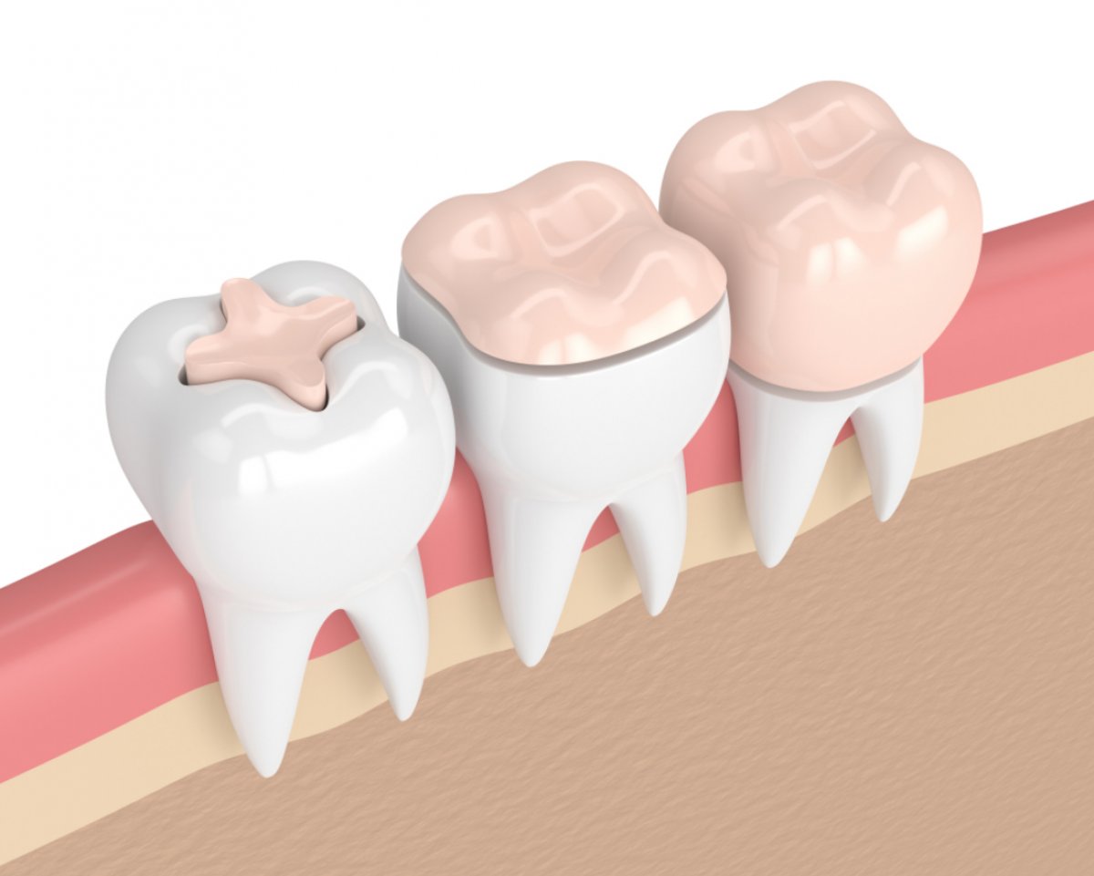
The success level of prosthetic and restorative applications performed on the oral and dental structure is directly related to the quality of the materials used and the precision with which the measurement stage is executed. This stage, forming the basis for procedures like porcelain veneers, zirconia crowns, E-Max restorations, or composite bonding, provides the necessary geographical map for the laboratory technician to achieve a millimetric fit without seeing the patient’s mouth. Innovative approaches in the industry, such as Aesthetic Dentistry, are integrating digital scanning technologies alongside manual methods into this process.
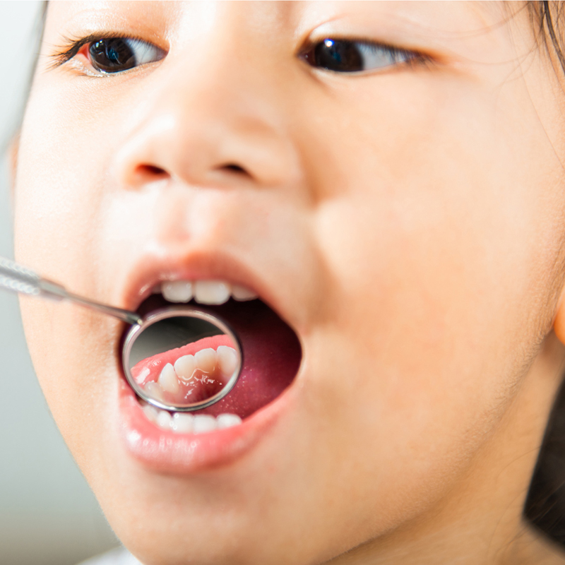
The measurement stage begins immediately after the minimal preparations or scanning preparations performed by the dentist on the tooth. The health of the gum tissue, the retraction cords prepared so that the measurement material can flow under the gum, and moisture control within the mouth constitute the technical details of this process. In practices conducted under the clinical standards of Avrupadent, digital scanning systems or traditional measurement trays are carefully selected with due consideration for the patient’s comfort.
Why is the measurement process in aesthetic dental treatment so important?
The only bridge between the laboratory and clinical practice in prosthetic dentistry is the impressions taken inside the mouth. A small mistake or millimetric deviation during the impression phase can lead to the porcelain produced in the laboratory being incompatible with the patient’s mouth, having a high profile, or leaving gaps. This situation prolongs the treatment duration and jeopardizes the health of the gums. Therefore, the dentist manages every detail, from the consistency of the impression material to the freezing duration inside the mouth, in accordance with protocols.
Especially in aesthetic work in the anterior region, the length, width, and distance from neighboring teeth of the tooth must be calculated at a micro level. The impression material must completely flow to the sulcus (pocket) surrounding the tooth, ensuring a clear depiction of the boundary where the porcelain will merge with the gum on the laboratory model. This precision is essential for achieving a flawless harmony in Aesthetic Dentistry practices.
What Materials and Trays Are Used in Traditional Impression Methods?
For years, the traditional method of taking impressions in dentistry has been carried out using two-stage or single-stage dual mix techniques. Initially, a general copy of the teeth is taken by placing silicone putty in the form of a thick paste. Then, low-viscosity liquid silicone is frequently injected around the teeth with special syringes by the dentist to capture the finest details (gingival margins), and the putty tray is reinserted into the mouth.
This process includes the waiting time of a few minutes required for the material to set in the patient’s mouth. Since the dimensional stability of the silicones used is quite high, they maintain their shape after setting, allowing the production of hard casting models when sent to the laboratory. In cases where traditional methods are preferred in Avrupadent clinics, a careful working protocol is followed to ensure that the material remains in a dry environment inside the mouth, eliminating the risk of deterioration.
How Does Digital Imprinting (Intraoral Scanner) Technology Work?
Intraoral scanners, one of the greatest revolutions in digital dentistry, create a colorful, three-dimensional, and highly detailed digital copy of the teeth and gums within seconds. As the dentist moves the tip of the scanner over the teeth, the camera instantly sketches a model on the screen. If there is an area that has not been scanned or is missing, the software immediately alerts the dentist, and the doctor rectifies the gaps by rescanning that area.
These digital data increase patient comfort because they do not require the freezing of physical measuring materials, providing great relief for individuals experiencing nausea reflex. The resulting three-dimensional digital model is transmitted to the laboratory or CAD/CAM milling devices within seconds via the internet. This rapid data flow ensures the acceleration of prosthetic production processes and minimizes the margin of error.
| Measuring Method | Materials / Tools Used | Patient Comfort and Process |
|---|---|---|
| Traditional Measurement | Silicone-based pastes/adhesive gels and trays | A few minutes of waiting in the mouth, sometimes a slight feeling of nausea. |
| Digital Measurement (Scans) | Optical camera (Intraoral scanner) and computer | Clean, fast, non-nauseating flow that can be controlled instantly. |
What Preparations Are Made in the Gingiva and Surrounding Tissues Before Taking Measurements?
Regardless of which measuring method is used, clearly seeing the boundary of the gingiva is essential for the edge adaptation of porcelain restorations. If there is active edema or bleeding in the gingiva, the measuring material cannot fully flow to that area, creating ambiguities in the laboratory model. Therefore, the physician ensures that the tooth cleaning or tissue regulation procedures are completed just before measurement.
Additionally, controlling moisture inside the mouth is particularly important in traditional silicone impressions. The covering of the outer surface of the impression prevents the material from adhering to the outside and capturing details. For this reason, cotton rolls, impression absorbers, or rubber-dam systems are used to isolate the working area and create a dry environment.
How Does Retraction Cord Clarify the Gum Margin?
The preparation boundaries for teeth to be covered with porcelain veneers or zirconium crowns are generally at the level of the gums or within the gum pocket (subgingival). It is difficult for these boundaries to exactly meet the impression due to the natural slight covering of the gums over the tooth. Cotton retraction cords impregnated with special medical formulations are the most common clinical tools used to ensure this margin is exposed.
The cord is placed around the tooth with special hand instruments without damaging the gum and is left for a few minutes. During this time, the cord gently separates the tissue, making the gum margin visible. While taking the impression, the cord is slowly removed and the fluid impression material is immediately injected into that space. This technique ensures the flawless replication of the margin that remains under the gum.
Challenges Encountered When Taking Impressions and How to Manage the Gag Reflex?
In some individuals, placing spoons or materials in the back of the mouth can trigger the gag reflex. This situation not only hinders the doctor from measuring comfortably but also creates stress for the patient. To overcome this difficulty in clinical practice, various medical and psychological approaches are employed. Patients are advised to breathe regularly and deeply through their noses to calm the reflexes in the soft palate area.
Furthermore, the use of digital scanners, which have much smaller tips compared to traditional measurement spoons, provides great comfort for patients with a gag reflex. Since no spoon is used in digital scanning, the process can be paused and restarted whenever needed, allowing patients to go through it much more comfortably and without stress.
How Are Taken Measurements Transferred to the Laboratory?
The acceleration of the prosthetic production phase is directly related to the speed of transferring measurement data to the laboratory. In traditional methods, after a silicone measurement is taken, it is disinfected and sent to the laboratory in special boxes. In the laboratory, a hard cast is poured inside this measurement to obtain a physical model of the patient’s mouth. On this cast, technicians produce porcelain using either wax modeling or digital scanning.
In digital workflows, courier processes are completely eliminated. The three-dimensional data set obtained with the intraoral scanner is instantly transferred in digital file format to the system of the Aesthetic Dentistry laboratory. The lab technician examines the model on the screen and starts the design process. This digital transfer prevents courier delays and makes it possible to start the production schedule much more quickly.
What Are the Differences Between Digital and Traditional Measurements?
Traditional silicone measurements can show variations on a micro level depending on the material’s setting time, room temperature, or the expansion rates during the pouring phase. However, digital measurements do not have the risk of physical deformation because optical scanners work with light waves. Whatever is displayed on the screen is exactly what the data sent to the laboratory is.
From the patient’s perspective, the pressure and taste sensations created by the material in the mouth are not present in digital scanning. From the doctor’s perspective, if an error is detected during scanning, it is sufficient to rescan only that area; it does not require starting the entire measurement from scratch. This efficiency is an important technological advantage that improves the quality of clinical workflow.
What Criteria Are Used for Temporary Restorations After Measurements?
After the preparations performed on the teeth during the porcelain laminate or zirconium veneer procedures, the production of permanent restorations in the laboratory may take a few days. During this waiting period, the exposure of the teeth can lead to stimulation of the dentinal tubules and cause severe sensitivity. To prevent this problem, temporary teeth made of acrylic or bis-acrylic composite materials are placed immediately after the impression session.
Temporary teeth, built with weak temporary cements, can be easily removed when the permanent teeth arrive. However, in this process, they play a critical role in protecting the health of the gum tissue and preventing the movement (shifting) of the teeth. In Avrupadent clinics, the contours of temporary restorations are meticulously prepared to not disturb the natural form of the gum tissue and meet aesthetic standards so that the patient can comfortably smile.
How Do Trials Conducted on the Model Affect the Treatment Flow?
After taking measurements and completing laboratory production, porcelain cannot be directly manufactured for teeth. A trial is first done in the model of the gum or in the patient’s mouth. The compatibility of zirconium frameworks and the occlusal balance between the upper and lower jaws are tested at this stage. If there is even the slightest incompatibility in the lengths of the teeth or contact points, it is reported to the laboratory for immediate revision.
This inspection stage serves as a safeguard against potential joint pain or porcelain fractures that may occur after the permanent manufacturing. The doctor also conducts phonetic sound tests (speech trials) at this stage to confirm that aesthetic and function are in harmony.
How Are Measurement and Digital Scanning Standards Applied in Avrupadent Processes?
In the institutional medical service standards, the success of prosthetic procedures begins with the importance given to the quality of measurements, which is the first step. In Avrupadent clinics, investments in digital infrastructures have been made in line with the goal of ensuring patient comfort and reducing the margin of error for the doctor to zero. Data obtained with optical scanners is transferred to the screens of expert technicians without any dimensional loss.
Doctors follow a meticulous protocol at every stage, from optimizing gum health to the compatibility of temporary teeth. These standards are the fundamental dynamics supporting the flawless fit of the produced porcelain laminates or zirconium crowns during the first trial in the patient’s mouth and their safe use for many years.
What Are the Frequently Asked Questions?
1. Will I feel pain or discomfort while taking the impression?
The impression-taking process is entirely painless; in traditional silicone impressions, there may be a temporary fullness in the mouth from the material and the spoon, while in digital scanning, only the contact of the camera is involved.
2. Which impression method is suitable for those with a gag reflex?
For individuals experiencing a strong gag reflex, a comfortable alternative is the digital intraoral scanning method, which works with a small scanner and can be paused at any time.
3. Which is more accurate, traditional or digital impressions?
Digital scanners provide superior accuracy compared to traditional silicone impressions because they operate with light waves and do not carry the risk of physical expansion/contraction, offering millimetric precision.
4. Will my teeth remain exposed after the impression is taken?
No, immediate placement of temporary teeth (crowns or veneers) after the impression session prevents sensitivity and maintains aesthetic appearance.
5. Why is dental gum retraction used and does it hurt?
It is used to clarify the gum line and ensure the complete acquisition of the impression; it may create a slight pressure sensation but does not cause pain due to the effect of local anesthesia.
6. How is digital impression data sent to the laboratory?
Without the need for physical courier, it is transmitted directly to the laboratory’s screen in seconds via cloud-based encrypted digital software.
7. How many days after the impression are the permanent teeth prepared?
The trials are generally completed within 5 to 10 days, depending on the density of the laboratory, the selected material (zirconium, E-Max, or laminate), and the number of teeth to be made.
8. What happens if there is a measurement error?
If there is even the smallest detail missing or a bubble is noticed in the measurement, the doctor immediately redoes the measurement to prevent compatibility issues with the porcelain.
9. Are temporary teeth placed immediately during the measurement session?
Yes, to protect your teeth while the laboratory preparation is ongoing after taking the measurement, temporary restorations are prepared and fitted within the same session.
10. Does taking the impression cause any problems?
Impression material can obstruct the adhesion of silicones and block the view of the digital camera; therefore, cotton and vacuums are used to keep the working area dry.
11. Does digital scanning emit harmful radiation?
No, intraoral scanners do not emit X-rays (radiation); they work completely harmlessly with optical light waves and camera technology.
12. Is sensitivity in my gums normal after the impression?
A slight sensitivity or tension feeling in the gum areas where the retraction cords are placed is considered normal within the first few hours and will pass in a short time.
13. Is taking a single tooth impression the same as taking an impression of the entire jaw?
Even when taking an impression for a single tooth, both upper and lower jaws must be fully measured or scanned to see the occlusion relationship with that tooth.
14. Can I eat after the impression is taken?
It is expected that the anesthesia will wear off after temporary teeth are placed; it is important to avoid very hard or sticky foods to prevent the temporary teeth from shifting.
15. Why is the measurement phase in European standards carried out with such precision?
Ensuring that porcelain restorations fit perfectly during the first trial in the mouth is managed with great precision to ensure durability and to protect the health of the gum tissue.