

How is the implant crown selected?
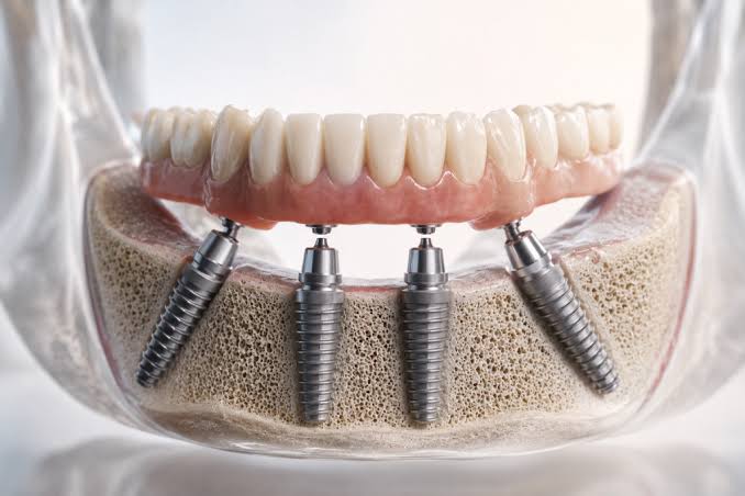
Surgical procedures performed to compensate for missing teeth complete the first medical phase with the osteointegration (integration with bone) process of the titanium artificial roots placed inside the jawbone. However, the primary structure that enables the patient to regain chewing, speaking (phonation), and aesthetic functions is the prosthetic restorations referred to as “implant crowns” that are fixed on these titanium roots. These restorations are made from inorganic materials that can withstand the high mechanical stresses produced by the jaw muscles and the biochemical environment of the oral cavity.
How is the implant crown selected and what criteria are considered?
There is no standard template in prosthetic dentistry; because each individual’s oral anatomy and chewing pattern (habit) is a unique engineering feat in itself. The shape and material of the tooth to be placed on the titanium root is shaped not only according to the patient’s cosmetic requests but also according to the distribution of the loads on the temporomandibular joint. When it is said, “how is the implant crown selected”, the first parameter looked at is the localization of the deficit.
In the visible areas where dental aesthetics are prioritized, high-translucency (light transmission) materials take precedence. The posterior areas (molars) are where the jaw muscles exert kilogram-level pressure, representing the areas where the gnashing process takes place. In these regions, the mechanical resistance (toughness) against breakage rather than the light permeability of the material is the main determinant in the dentist’s decision-making process. Additionally, whether the teeth opposing the patient’s jaw are natural or prosthetic is among the factors that determine the level of aesthetics for the selected material.
What Are the Types of Materials for Implant-Supported Dental Treatment?
Each material prepared in a laboratory has its own physiological advantages and biomechanical limits. Dentists process these materials in special furnaces at high temperatures (approximately 1000°C) to transform them into glassy and non-porous forms according to the measurements of the patient. The fundamental purpose of the materials is to mimic the physical properties of natural enamel and dentin at a medical level.
- Zirconium Dioxide (ZrO2): A white-colored alloy that possesses both high mechanical strength and aesthetic light transmission. It has high tissue compatibility due to the absence of metal.
- Metal Framework Porcelains: These are traditional structures that contain a durable metal framework, such as chrome-cobalt, in their inner part and are covered with aesthetic ceramic on the outer surface. Their fracture resistance is very high.
- Lithium Disilicate (Full Ceramics): Structures completely made of glass ceramics, which have no supporting framework in their infrastructure, are very similar in optical reflection to natural teeth but have somewhat limited fracture resistance compared to zirconia.
In Which Situations Are Zirconium Implant Crowns Preferred?
In dentistry, aesthetics is measured by how much visual illusion is created in harmony with surrounding tissues, rather than just how white a tooth is on its own. Zirconium is one of the most modern restorative materials that support this illusion. Traditional metals block the light that falls on them, causing the tooth to appear lifeless and dull; whereas zirconium allows the light to enter the prosthetic body and reflect. This situation helps achieve an indistinguishable harmony with natural teeth right next to the restoration.
In addition to aesthetic contributions, the physiological interaction established by zirconium with the mucosa of the dental tissue is clinically quite significant. On highly polished zirconium surfaces, the adhesion and organization of bacterial plaque (biofilm) is very challenging. Limiting plaque accumulation helps reduce the risk of inflammation (peri-implantitis) around the titanium root, thereby maintaining the long-term health of the system. Lack of metal ions also prevents gray-purple discolorations that may develop over time at the gum margin.
Who is Suitable for Metal Supported Porcelain Implant Crowns?
The back areas of the human jaw (molar tooth areas) are subjected to kilograms of pressure due to the contraction of the masseter and temporal muscles. The primary focus for the clinician during prosthetic planning in these areas is to limit the potential for the restoration to break or bend (fracture). Metal framework porcelains, due to the biocompatible metal alloys they contain (usually cobalt-chromium or nickel-chromium), are the materials with the highest resistance to bending.
Especially in anatomical cases where three or more adjacent teeth are missing and long-bodied bridges will be placed between the titanium roots, any pressure exerted on the body section tries to buckle the bulk material. The metal framework acts as a structural scaffold against this buckling moment. Aesthetically, while it has a certain opacity due to the metal inside, this does not pose a medical disadvantage as it remains out of the visual field in the back areas.
What to Consider in Choosing Anterior Implants?
The anterior incisors and canine (dog tooth) areas included in the smile line are the most anatomically sensitive areas in prosthetic dentistry. In these areas, the gum (mucosa) structure generally has a thin biotype. If a metallic connecting piece (abutment) is placed on a titanium root and a thick prosthetic structure is placed on top, the cold gray color of the metal may shine through the thin gum tissue.
Doctors plan to use “Zirconium Abutments” instead of standard titanium abutments in the anterior area to overcome this anatomical handicap. When zirconium pieces are cemented (constructed) with either full ceramic or zirconium crowns, the entire system gains a white-transparent form. When light hits the tooth, it descends to the tooth root without hitting a metal barrier and presents a lively, pink, and homogeneous appearance as if a natural tooth is emerging from there.
Which Implant Supported Teeth Are Planned for Posterior Chewing Teeth?
The biomechanical environment in the posterior dental regions is quite vulnerable to parafunctional (abnormal) muscle movements such as teeth grinding (bruxism) or teeth clenching. If a patient tightly grinds their teeth during sleep at night, it is crucial that the material chosen for the upper structure does not break down under these horizontal (shearing) forces. While porcelain materials are resistant to compressive (vertical) forces, they may experience minor surface fractures known as “chipping” under shearing forces.
To eliminate these medical risks, the use of “Monolithic Zirconium” (Single Piece Zirconium) has become quite common in recent years for posterior areas. In monolithic restorations, there is no metal framework or porcelain layer; the entire tooth is obtained from a single zirconium block using digital milling devices. Because it has a monolithic structure, the risk of porcelain detachment (delamination) is biophysically eliminated, and chewing mechanics can be maintained for many years.
Are Implant Overdentures Fixed with Screws or Cemented?
The connection of the prosthetic superstructure to the titanium artificial root is an important junction where medical engineering comes into play in dentistry. Both stabilization methods have physiological advantages depending on the patient’s anatomy and ease of use.
| Parameter | Vidalı Sabitleme (Screw-Retained) | Yapılandırma (Simante) Sabitleme |
|---|---|---|
| Mekanik Uygulama | The prosthesis is screwed directly into the underlying titanium root with a micro screw that passes through the chewing surface. | An abutment is screwed onto the titanium root, and the prosthesis is constructed over this piece using medical cements. |
| Klinik İdame (Bakım) | The doctor can remove the filling from the screw socket at any time to take out the prosthesis, clean it, and put it back in. The maintenance is quite easy. | Removing the constructed prosthesis is difficult. The prosthesis may get damaged during the removal process. |
| Estetik ve Açık | A closed screw hole is present on the chewing surface. If the titanium root fixture is placed, the hole may coincide with the visible anterior surface, negatively affecting aesthetics. | There is no screw hole; it is completely in the natural tooth form. The titanium roots offer flexible use to compensate for open defects. |
| Biyolojik Risk | Since there is no structure that would leak under the gum, the risk of peri-implantitis (tissue inflammation) is more limited. | If the remnants of the structure (cement) are not completely cleaned from the gum pocket, it may initiate inflammation that leads to bone resorption in the area. |
How is Implant Overdentistry Planned in İzmir Avrupadent?
In corporate medical organizations, the process is not just a one-dimensional task like taking measurements from the patient’s mouth and sending them to the laboratory. The success of the restoration is dependent on the ‘Backward Planning’ protocol, which starts before the surgical phase. In this protocol, before deciding where to place the titanium screw in the bone, the doctor completes the 3D design of the tooth (crown) that will come to that area in the virtual environment.
Once the dimensions of the prosthesis, the relationship with adjacent teeth, and the chewing center are determined, the titanium screw is positioned in the computer environment to exit from directly beneath this center. This analytical approach ensures that the artificial root shares the same mechanical axis with the prosthesis above it. Thus, off-axis forces are prevented, which medically restricts the tensile forces on the titanium neck and prevents bone resorption.
How is the Implant Crown Color Matched to Natural Teeth?
Color detection is one of the most delicate aspects of medical aesthetics. A human tooth is not made up of a single, homogeneous shade; the neck area (the area close to the gum) exhibits a slightly yellower dentin reflection, while as we move towards the cutting edges, the enamel layer becomes more transparent, resulting in bluish/greyish light permeability (opalescence). A prosthesis produced in a plain and pure white color is immediately noticeable as an opaque and artificial block among the adjacent teeth.
Dentists map not only the primary color (Hue) when determining the color of restoration but also the saturation (Chroma) and the degree of brightness/grayness (Value). Microscopic cracks, white spots (hypocalcifications), or small yellows on the tooth surface can be transferred to the laboratory technician using digital photos and polarized filters. The technician layers the porcelain powders onto the crown using a brush in a layer-by-layer manner (layering technique), thus precisely replicating the biological anatomy.
How Do Digital Measurement Systems Work in Implant-Supported Tooth Restorations?Digital measurement systems swiftly create a three-dimensional map of the jaw within seconds using intraoral optical cameras, eliminating the gag reflex caused by silicone measurement materials. The digital data obtained with specially designed scanning parts placed over a titanium root is transferred to CAD/CAM software, paving the way for the production of implant-supported dental prostheses with millimetric accuracy.
In traditional measurement methods, trays filled with paste-like materials (alginate or silicone) are placed in the patient’s mouth, and it would take a few minutes for the material to set. During this time, the material could deform, or the patient’s oral isolation could be disrupted, leading to micron-level deviations in the measurement. In digital dentistry, pen-sized intraoral (inside the mouth) scanners are used.Medical Advantages of Digital Workflow:- Micron Level Precision: Geometric parts known as “Scan Body” are attached to the titanium root. The scanner perfectly detects the position and angle of this part, accurately calculating the marginal boundary (the edge sealing) where the prosthesis will sit on the root.
- Time Savings: Measurements are instantly sent to the laboratory via cloud systems; by limiting the waiting times for impressions and models, the production schedule is narrowed.
- Digital Archiving: The dynamics of the patient’s jaw closure are stored in a digital environment. If a fracture occurs in the prosthesis in the coming years, the same prosthesis can be retrieved within minutes from milling devices using the archived data without the need to take new measurements from the patient.
How Does Jaw Closure (Occlusion) Affect the Selection of Implant-Supported Teeth?
Para-functional habits such as the patient’s jaw closure relationship (occlusion) and teeth grinding (bruxism) directly shape the choice of materials. In individuals with high chewing pressure, implant-supported dental blocks with high toughness, such as monolithic zirconium, are designed to prevent porcelain chipping, with the goal of transmitting mechanical forces to the bone in a physiological balance.In jaw anatomy, the way the lower and upper teeth contact each other (occlusion) is the safeguard of the biomechanical system. Natural teeth are connected to the jawbone by flexible “periodontal ligament” fibers. These fibers allow the tooth to slightly flex within the bone (shock absorption) when excessive pressure occurs. However, titanium roots are directly integrated into the bone (in the form of ankylosis); they have no room for flexing.
Due to this rigid (non-flexible) structure, if the prosthesis contacts the opposing tooth surface by even half a millimeter (early contact), all chewing forces hit a single point like a hammer. This destructive stress can either crack the porcelain on the prosthesis or erode the jawbone around the titanium root. To prevent this situation, dentists control the prostheses with articulation papers of micron thickness. Additionally, if the patient is diagnosed with severe teeth grinding (bruxism), zirconium or hybrid restorations that do not carry a risk of breakage are planned instead of using porcelain coating.
How Should the Cleaning and Maintenance of Implant-Supported Dental Prostheses Be Done?
In addition to regular tooth brushing, it is necessary to wash these prostheses daily using specialized brushes and superfloss threads to clean the narrow gaps between the titanium neck region and the gum. Professional tartar cleaning performed during periodic dental examinations helps to limit potential inflammations in surrounding tissues and protects the integrity of the material.Since the prostheses produced in the laboratory have an inorganic structure, they are not affected by microbial acids. However, the jawbone and mucosa (gum) under the prosthesis are living tissues. If the patient neglects oral hygiene, food residues and salivary enzymes can form a layer of “biofilm” (bacterial plaque) around the neck of the prosthesis. Plaque that is not cleaned hardens into dental calculus (tartar) within twenty-four hours.
Given that the processed dental implants have a porous surface, they create safe havens for bacteria. These bacteria can migrate down from the gum tissue and initiate a bone resorption inflammation known as “peri-implantitis” around the titanium root. Cleaning the cavities underneath the prostheses (especially in bridge designs involving multiple teeth) with pressurized water flossers in situations where mechanical toothbrushes cannot reach is the most favorable medical maintenance method that disrupts the colonization of these harmful bacteria that thrive in anaerobic environments.
Which Implant Surface Dental Material Stands Out in Terms of Biocompatibility?
The material that provides the highest adaptation to tissues from a biocompatibility perspective is zirconium. Zirconium implant surface dental restorations, with their smooth surface that minimizes plaque retention and inorganic structure that does not contain metal, are quickly accepted by gum cells, thereby limiting allergic reactions or gray color reflections at the mucosal level.Biocompatibility is the ability of a foreign substance placed in the oral environment to establish physiological harmony without causing any toxic, inflammatory, or allergic reaction in local tissues. Metals like nickel or cobalt, typically used in the frameworks of traditional porcelains, can oxidize (corrode) over time by exchanging ions with oral fluids. This oxidation can lead to dark gray discolorations (metallic margins) resembling tattoos in the gums.
Zirconium (zirconium dioxide), like titanium, is a tissue inert (non-reactive) ceramic. Gum cells (fibroblasts) exhibit a high affinity towards the smooth surface of zirconium, effectively enveloping the material as if it were their own enamel tissue. This tight mucosal barrier serves as a physiological seal that mechanically blocks food residues and bacteria from seeping beneath the titanium root.
What Factors Play a Role in Implant-Supported Dental Applications in the İzmir Region?
Izmir’s advanced medical infrastructure and innovative clinical protocols allow for the progression of implant-supported dental design processes in accordance with the standards of digital dentistry. The coordinated collaboration of oral surgeons and prosthodontists ensures the planning of physiological restorations suitable for the patient’s facial anatomy, supporting the long-term medical maintenance of biomechanical balance.In regions where technology and specialization are integrated in the healthcare field, prosthetic treatments evolve from a “product delivery” to a sustainable “tissue rehabilitation” process. In the İzmir location, corporate organizations that establish a European-style medical ethics center operate specific clinical algorithms that blend material science with the hospital’s cellular capacity.
In traditional measurement methods, trays filled with paste-like materials (alginate or silicone) are placed in the patient’s mouth, and it would take a few minutes for the material to set. During this time, the material could deform, or the patient’s oral isolation could be disrupted, leading to micron-level deviations in the measurement. In digital dentistry, pen-sized intraoral (inside the mouth) scanners are used.
- Micron Level Precision: Geometric parts known as “Scan Body” are attached to the titanium root. The scanner perfectly detects the position and angle of this part, accurately calculating the marginal boundary (the edge sealing) where the prosthesis will sit on the root.
- Time Savings: Measurements are instantly sent to the laboratory via cloud systems; by limiting the waiting times for impressions and models, the production schedule is narrowed.
- Digital Archiving: The dynamics of the patient’s jaw closure are stored in a digital environment. If a fracture occurs in the prosthesis in the coming years, the same prosthesis can be retrieved within minutes from milling devices using the archived data without the need to take new measurements from the patient.
How Does Jaw Closure (Occlusion) Affect the Selection of Implant-Supported Teeth?
In jaw anatomy, the way the lower and upper teeth contact each other (occlusion) is the safeguard of the biomechanical system. Natural teeth are connected to the jawbone by flexible “periodontal ligament” fibers. These fibers allow the tooth to slightly flex within the bone (shock absorption) when excessive pressure occurs. However, titanium roots are directly integrated into the bone (in the form of ankylosis); they have no room for flexing.
Due to this rigid (non-flexible) structure, if the prosthesis contacts the opposing tooth surface by even half a millimeter (early contact), all chewing forces hit a single point like a hammer. This destructive stress can either crack the porcelain on the prosthesis or erode the jawbone around the titanium root. To prevent this situation, dentists control the prostheses with articulation papers of micron thickness. Additionally, if the patient is diagnosed with severe teeth grinding (bruxism), zirconium or hybrid restorations that do not carry a risk of breakage are planned instead of using porcelain coating.
How Should the Cleaning and Maintenance of Implant-Supported Dental Prostheses Be Done?
Since the prostheses produced in the laboratory have an inorganic structure, they are not affected by microbial acids. However, the jawbone and mucosa (gum) under the prosthesis are living tissues. If the patient neglects oral hygiene, food residues and salivary enzymes can form a layer of “biofilm” (bacterial plaque) around the neck of the prosthesis. Plaque that is not cleaned hardens into dental calculus (tartar) within twenty-four hours.
Given that the processed dental implants have a porous surface, they create safe havens for bacteria. These bacteria can migrate down from the gum tissue and initiate a bone resorption inflammation known as “peri-implantitis” around the titanium root. Cleaning the cavities underneath the prostheses (especially in bridge designs involving multiple teeth) with pressurized water flossers in situations where mechanical toothbrushes cannot reach is the most favorable medical maintenance method that disrupts the colonization of these harmful bacteria that thrive in anaerobic environments.
Which Implant Surface Dental Material Stands Out in Terms of Biocompatibility?
Biocompatibility is the ability of a foreign substance placed in the oral environment to establish physiological harmony without causing any toxic, inflammatory, or allergic reaction in local tissues. Metals like nickel or cobalt, typically used in the frameworks of traditional porcelains, can oxidize (corrode) over time by exchanging ions with oral fluids. This oxidation can lead to dark gray discolorations (metallic margins) resembling tattoos in the gums.
Zirconium (zirconium dioxide), like titanium, is a tissue inert (non-reactive) ceramic. Gum cells (fibroblasts) exhibit a high affinity towards the smooth surface of zirconium, effectively enveloping the material as if it were their own enamel tissue. This tight mucosal barrier serves as a physiological seal that mechanically blocks food residues and bacteria from seeping beneath the titanium root.
What Factors Play a Role in Implant-Supported Dental Applications in the İzmir Region?
In regions where technology and specialization are integrated in the healthcare field, prosthetic treatments evolve from a “product delivery” to a sustainable “tissue rehabilitation” process. In the İzmir location, corporate organizations that establish a European-style medical ethics center operate specific clinical algorithms that blend material science with the hospital’s cellular capacity.







