

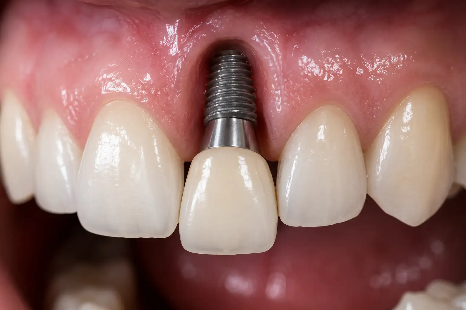
Is a gum graft necessary for an implant?
In the restoration of tooth deficiencies, the focus is often shifted to the volume of the jawbone and the biological integration (osteointegration) of titanium materials with the bone. However, when evaluated in light of medical engineering and physiological principles, the real barrier that protects the jawbone and the artificial material within it from microbial agents in the external environment is the gum tissue (mucosa) that covers the bone. In cases where the structure of the gum is thin, sensitive, or tense, the underlying bone tissue remains unprotected against external factors. At this point, the disciplines of jaw surgery and periodontology (gum diseases) come together to develop medical strategies aimed at increasing tissue thickness.
When is a gum graft needed for an implant and under which anatomical conditions is it planned?If the gum tissue in the area where titanium artificial roots will be placed is structurally very thin, is of a flexible mucosa type that is not resistant to chewing forces, or if the bone surface has remained unprotected due to past tissue pullbacks, performing a gum graft for the implant is a medical necessity. This surgical procedure aims to thicken the soft tissue in the area to create a biological seal (barrier) against bacterial attacks.
In dental practice, when tooth deficiency occurs in an area, not only does the underlying jawbone (alveolar bone) lose volume; the gum that covers that bone also becomes dysfunctional and shrinks to assume a thin form due to its ineffectiveness. When titanium materials are placed into the bone, there is a fine boundary where the neck parts are exposed to the oral environment. The quality of the gum surrounding this boundary is the only anatomical shield that ensures the system remains free from infection for many years.If the gum in this area is as thin as the thickness of a razor or if it has an elastic structure that can easily shift with lip-cheek movements, during brushing or chewing, this tissue may detach from the titanium neck. Food remnants and pathogenic bacteria that seep in through the microscopic gaps can directly reach the bone, leading to a condition called “peri-implantitis,” which initiates inflammatory bone resorption. The gum graft (soft tissue transplant) procedure is planned precisely to address this anatomical weakness, thicken the tissue, and create a resilient mucosal band against physical trauma from the outside.
What is Keratinized Gum and Why is it Crucial for Titanium Roots?Keratinized gum is a thick mucosal tissue that tightly surrounds the necks of natural teeth and titanium roots, resistant to the abrasions encountered during chewing, immobile, and pale pink in color. For the long-term maintenance of titanium materials, there must be at least a 2-millimeter wide band of keratinized gum around them; this is essential to limit bacterial seepage, tissue retraction, and local inflammations.
When we examine oral anatomy, we encounter two different types of soft tissue. The first is the “alveolar mucosa,” which covers the inner surface of the cheeks, the underside of the lips, and the floor of the mouth. This tissue is reddish, very thin, flexible, and mobile. The second is the “keratinized gum,” which covers our palate and is located immediately around the teeth. This tissue is tightly attached to the skeleton, hard, immobile, and acts almost like armor against mechanical trauma.Titanium roots are placed into the bone and porcelain teeth (crowns) are added on top. During the brushing of these artificial teeth, the bristles come into contact with the boundary between the tooth and the gum. If at that boundary, instead of firm and resilient keratinized gum tissue, flexible alveolar mucosa is present, every contact of the toothbrush with that area leads to irritation, exudation, and micro-bleeding. As the individual cannot brush comfortably, plaque accumulation in that area increases. To break this physiological reaction chain, doctors transplant hard connective tissue taken from the palate to the thin mucosal area to convert it into a keratinized band.
How is the Decision for Gum Graft Taken at Avrupadent Clinics in Izmir?
In multidisciplinary medical organizations like Avrupadent, which provide services in Izmir, the decision for gum grafting is shaped by clinical examinations performed by periodontology (gum diseases) and jaw surgery specialists. The thickness of the gum (biotype) is measured with medical probes, the pulling forces of the cheek muscles are evaluated, and when an anatomical deficiency is detected, a soft tissue transplant protocol is integrated into the surgical calendar.The institutional medical understanding does not see interventions in the jawbone merely as a “bone inside” procedure. While three-dimensional dental tomographs (CBCT) provide millimetric measurements of the bone, the millimetric thickness of the gum can only be determined through the doctor’s physical examination and special periodontal instruments. If the doctor applies a slight force horizontally to the gum in the relevant area and the tissue moves freely, a diagnosis of insufficient stable connective tissue is made.
Criteria for Evaluating Gum Biotype:- Thick Biotype: The gum tissue is bulky, fibrotic (stiff), and resilient. It masks the gray color of the underlying titanium. Usually, an additional graft procedure is not necessary.
- Thin (Scalloped) Biotype: The gum tissue is as thin as paper, turns red. When titanium is placed or when a porcelain crown is placed later, it easily shows recession tendencies. Grafting becomes a clinical necessity for individuals with this biotype.
- Frenulum Tension: If the muscle attachments (frenulum) that connect the lips and cheeks to the gum tissue are positioned very high, they pull the gum tissue downward during speaking. To limit this pulling force, the attachments are released and a graft is added in between.
How is Gum Grafting (Soft Tissue Grafting) Performed Using Medical Methods?
Soft tissue grafting is typically performed by securing a piece of connective tissue or epithelial tissue taken from the patient’s own palate (donor site) to the deficient thin gum area (recipient site) using microsurgical stitches. Depending on the need, the physician selects the most suitable technique from Free Gingival Graft (FGG) or Connective Tissue Graft (CTG) techniques according to the anatomy.This surgical intervention is entirely based on the principle of autograft (transfer of the individual’s own tissue). Since the body’s own tissue is used, no cellular rejection reaction is expected. The technique to be applied may vary according to aesthetic expectations and the type of tissue needed.
Surgical Techniques for Soft Tissue Grafting Surgical Method Clinical Procedure Medical Application Area Free Gingival Graft (FGG) The surface of the palate should be taken as a segment, along with the outer layer (including epithelium). Especially in the back areas of the lower jaw, it is planned primarily to form a keratinized (hard) tissue band rather than for aesthetic purposes. The color may heal in a slightly lighter tone compared to the surrounding tissue. Connective Tissue Graft (CTG) The outer layer (epithelium) of the palate is lifted like a flap, and only the underlying connective tissue is taken. The outer flap is then closed back. In the anterior (gingival line) areas, it is used to increase volume and mask the gray reflection of titanium. The color harmony with the surrounding tissue is perfect.
Can Soft Tissue Grafting Be Done in the Same Session as Implant Placement?
If the bone volume and blood supply in the recipient area are suitable, the implant placement with titanium and the gum graft can be integrated and performed in the same surgical session. This simultaneous procedure supports the patient to go through a single surgical recovery phase, maximizing time efficiency. However, in complex cases with severe tissue deficiency, the graft procedure can be planned as a separate session months before the titanium placement.In medical planning, time management is shaped by the biological tolerance of the tissues. On the day when the titanium material is placed in the jawbone, the gum is already surgically opened. The surgeon can position the connective tissue taken from the palate like a cushion inside the opened (flap lifted) gum tissue and stitch it shut. This procedure, performed simultaneously, allows for the formation of a thick mucosal barrier around the titanium implant right from the start.
On the other hand, if there is no keratinized gum tissue left in that area or if the lip muscles excessively retract the area, the doctor may prefer to graft that area first and allow it to heal. After an approximately eight-week cellular maturation (vascularization) period, a healthy, thick, and immobile tissue band will form in that area. In the second session, a titanium screw is passed through this reinforced safe tissue and placed into the bone. Both approaches are in accordance with medical norms; the decision is determined by the patient’s existing tissue map.
What is the Healing Process in the Donor (Palate) and Recipient Area After Surgery?
In the days following the operation, a slight tissue exudate and fine whitening associated with cellular repair in the donor area, which is the palate, is a physiological reaction. In the recipient area where the graft is placed, an average biological integration phase of two to four weeks occurs for the establishment of capillary vascularization (angiogenesis); this process is managed at normal levels with the pain relievers and protective plates prescribed by the doctor.Patients who have undergone soft tissue surgery typically feel slight sensitivity in the palate area from which the graft is taken, rather than in the bone area where the titanium material is placed. The palate is a highly vascularized area and has a high self-renewal rate. In the connective tissue graft (CTG) technique, there is only a fine incision line in the palate, and since it is covered with stitches, healing quickly improves within a few days.
When the free gingival graft (FGG) is applied, a superficial and open wound area forms on the palate (similar to a scratch on the skin). To ensure that this open area does not discomfort the patient, special transparent protective plates (Essix) or tissue stabilizers are used to cover that area at the end of the operation. In the recipient area where the transplant is performed, it is essential for the graft to sustain its vitality by nourishing from the surrounding tissues. In the initial days, the graft may appear whitish or pale, but as new capillaries progress into it (vascularization), it will return to a pink and vibrant form.
What Risks Does the Application of Titanium Material Pose in Areas with Gum Recession?
Placing titanium material in areas with gum recession and where the mucosal barrier has thinned, without performing tissue thickening, exposes the tissue to bacterial infiltration, risks the titanium’s gray-metallic reflection being visible externally, and creates risks for progressive bone resorption (peri-implantitis) due to mechanical irritation during brushing.The titanium screws placed into the jawbone have a grayish metal color as required by nature. No matter how aesthetic the zirconium or porcelain teeth placed on them are, the transition point between the tooth and the gum is a critical threshold. In individuals with gum recession, this boundary tissue is tissue-paper thin. When light passes through this thin tissue, it reflects the underlying metallic color, causing the gums to appear purple, gray, or lifeless.
Beyond the aesthetic issue, the real medical danger is the collapse of the biological defense. A retracted and thin gum loses its elasticity. When the patient brushes that area, the thin mucosa may tear or get irritated. In the irritated area, the patient stops brushing; plaque accumulates where brushing has stopped. The bacteria within the plaque reach the jawbone directly, as they do not encounter any physical barrier (thick mucosal band), leading to tissue destruction. A gum graft breaks this vicious cycle, providing both aesthetic permanence and medical protection to the tissue.
What Should Patients’ Nutrition and Hygiene Routines Be After Surgery?
During the first two weeks following the graft procedure, it is essential to completely isolate the transplant area from mechanical movement and to consume foods at room temperature in liquid or puree consistency. It is important to avoid brushing the relevant area with a toothbrush; plaque control should be ensured with chlorhexidine-based mouth rinses as recommended by the physician to support the drying of the capillary vessel connections of the transplanted tissue.The most critical factor determining the success of soft tissue grafts is “stabilization.” The tissue piece obtained from the palate does not have its own vascular system; it requires new capillary vessels that will reach out from the underlying bed tissue (the recipient area) to survive. If the patient chews a hard morsel or brushes the area vigorously in the first few days, the graft may shift millimetrically. Since the capillary vessels are still very fragile, this micro-mobility can break the vessels, leading to the graft not being nourished and resulting in death (necrosis).
Recommendations for Medical Healing Period:- Mechanical Protection: During the first 14 days, no chewing should be done in the relevant area. One should avoid facial expressions that overly stretch the lips (smiling, yawning) and refrain from pulling the lip with the hand to check the tissue.
- Dietary Discipline: Hot foods can dilate blood vessels and trigger bleeding. Cold foods may slow down circulation. Foods should be at room temperature (warm) and non-acidic.
- Chemical Hygiene: In this region where the use of toothbrushes is prohibited, to limit bacterial load, chemical antiseptic mouthwashes should be passively held in the mouth twice a day (without vigorous rinsing) and should be slowly expelled.
Can Gingival Graft Surgery Be Planned for Individuals in the Elderly Age Group?
In individuals in the elderly (geriatric) group, chronological age does not constitute an obstacle; however, the overall systemic health status and cellular perfusion capacity should be taken into account. Surgical graft procedures can be applied within medical norms to aging individuals whose medical tests (blood sugar, clotting profile) are within reference ranges, as long as surgical trauma is kept to a minimum.There is a misconception in society that soft tissue surgeries are only performed on young individuals concerned about aesthetics. However, with the aging process, a natural thinning (atrophy) and retraction in the gums is a physiological reality. The biological defense of titanium roots placed in the jaws of elderly individuals is more fragile due to this thin tissue.
Doctors are careful not to prolong the procedure while planning grafts in geriatric patients and try to keep the wound area in the palate as small as possible. A slight slowdown in cellular renewal speed due to aging (delayed wound healing) is taken into consideration. The anticoagulant agents used are adjusted under the supervision of the doctor to ensure the safety of the operation. Achieving a healthy mucosa is an important medical investment for protecting the individual’s prosthesis from environmental infections for many years.
How Do Systemic Diseases and Tobacco Use Affect Gum Healing?
Chronic diseases such as diabetes and tobacco use weaken the capillary circulation (microvascularization) within the mucosa, disturbing the nourishment of the graft. Since the vasoconstrictive effect of nicotine will limit the arrival of repair cells to the area, it is essential to maintain metabolic balance and limit tobacco consumption before surgical procedures in individuals with such risk factors.Gum tissue transplantation is a tissue engineering procedure entirely based on vascularization (blood supply). In uncontrolled Type 2 diabetes patients, high blood glucose damages the proteins in the vessel walls, limiting the oxygenation of the tissues. A non-nourished graft piece cannot survive and is expelled from the area as infected. Therefore, in diabetic patients, the decision for surgery is made after HbA1c levels are pulled within reference ranges.
The use of tobacco is the most significant enemy of wound healing. Cigarette smoke creates direct heat and toxic chemical accumulation in the oral mucosa. While nicotine narrows the blood vessels that carry oxygen to the tissues (vasoconstriction), carbon monoxide binds to oxygen in the blood, causing cellular disruption. Oral surgeons report that patients undergoing gum grafts should stop using tobacco at least two weeks before the operation and restrict its use during the initial months after the cellular integration is complete as a medical rule.
Why is Gum Graft Applied in Areas with High Aesthetic Expectations (at the Smile Line)?
Gum grafts are applied to mimic the natural form of pink aesthetics (gum) surrounding white aesthetics (zirconium crown) in cases of anterior tooth deficiencies. This transplant procedure increases the thickness of the gum, blocks the gray reflection of titanium, creates a symmetric gum line (crest line) with neighboring teeth, and supports the fullness of the gum papillae (triangular tissues).Gum aesthetics are evaluated not only by the color of the produced porcelain or zirconium tooth but also by how naturally it emerges from the gum (emergence profile). When a tooth extraction occurs in anterior regions (maxillary anterior), the surrounding bone and gum rapidly exhibit a tendency to shrink. When a titanium screw is placed in a shrunk area, even if covered with porcelain, the gum line remains higher than that of neighboring teeth, resulting in an asymmetric (long) dental appearance.
The connective tissue grafts applied to solve this aesthetic and anatomical issue essentially fill the recessed area from within, gaining volume. Doctors manipulate this transplanted thick tissue with temporary prostheses (gum reshapers) to ensure that the porcelain crown is enveloped in a natural wave form. Thus, the final restoration creates a perfect visual illusion, anatomically resembling the original tooth.
Why Should Long-Term Post-Surgical Oral Care and Clinical Maintenance Appointments Not Be Missed?
The long-term stability of the mucosal band obtained through graft surgeries and the underlying titanium structure is dependent on the removal of microbial plaques before they can organize. In addition to individual mechanical brushing and device cleaning, professional clinical maintenance sessions conducted by specialist physicians every six months provide medical support to prevent cellular damage.The anatomical integrity obtained through gum surgeries and titanium root applications is a dynamic system that requires maintenance. The idea that “the tissue has thickened, so there are no more issues” is not compatible with physiological cycles. No matter how thick and resilient the mucosa around the titanium is, if the patient neglects daily hygiene routines (brushing, using superfloss, and mouthwash), the plaques that accumulate in that area will eventually turn into tartar (calculus).
The porous surface of dental tartar creates a sheltered environment for bacteria, triggering gum inflammation. In routine check-ups every six months, doctors remove the invisible plaques in these blind spots (within the sulcus) using Teflon or carbon curettes that do not damage the titanium surface. Simultaneously taken radiographic images monitor the stability at the bone level. This periodic medical follow-up is the only guarantee that the biomechanical system will function healthily for decades.
In dental practice, when tooth deficiency occurs in an area, not only does the underlying jawbone (alveolar bone) lose volume; the gum that covers that bone also becomes dysfunctional and shrinks to assume a thin form due to its ineffectiveness. When titanium materials are placed into the bone, there is a fine boundary where the neck parts are exposed to the oral environment. The quality of the gum surrounding this boundary is the only anatomical shield that ensures the system remains free from infection for many years.
If the gum in this area is as thin as the thickness of a razor or if it has an elastic structure that can easily shift with lip-cheek movements, during brushing or chewing, this tissue may detach from the titanium neck. Food remnants and pathogenic bacteria that seep in through the microscopic gaps can directly reach the bone, leading to a condition called “peri-implantitis,” which initiates inflammatory bone resorption. The gum graft (soft tissue transplant) procedure is planned precisely to address this anatomical weakness, thicken the tissue, and create a resilient mucosal band against physical trauma from the outside.
What is Keratinized Gum and Why is it Crucial for Titanium Roots?Keratinized gum is a thick mucosal tissue that tightly surrounds the necks of natural teeth and titanium roots, resistant to the abrasions encountered during chewing, immobile, and pale pink in color. For the long-term maintenance of titanium materials, there must be at least a 2-millimeter wide band of keratinized gum around them; this is essential to limit bacterial seepage, tissue retraction, and local inflammations.
When we examine oral anatomy, we encounter two different types of soft tissue. The first is the “alveolar mucosa,” which covers the inner surface of the cheeks, the underside of the lips, and the floor of the mouth. This tissue is reddish, very thin, flexible, and mobile. The second is the “keratinized gum,” which covers our palate and is located immediately around the teeth. This tissue is tightly attached to the skeleton, hard, immobile, and acts almost like armor against mechanical trauma.Titanium roots are placed into the bone and porcelain teeth (crowns) are added on top. During the brushing of these artificial teeth, the bristles come into contact with the boundary between the tooth and the gum. If at that boundary, instead of firm and resilient keratinized gum tissue, flexible alveolar mucosa is present, every contact of the toothbrush with that area leads to irritation, exudation, and micro-bleeding. As the individual cannot brush comfortably, plaque accumulation in that area increases. To break this physiological reaction chain, doctors transplant hard connective tissue taken from the palate to the thin mucosal area to convert it into a keratinized band.
How is the Decision for Gum Graft Taken at Avrupadent Clinics in Izmir?
In multidisciplinary medical organizations like Avrupadent, which provide services in Izmir, the decision for gum grafting is shaped by clinical examinations performed by periodontology (gum diseases) and jaw surgery specialists. The thickness of the gum (biotype) is measured with medical probes, the pulling forces of the cheek muscles are evaluated, and when an anatomical deficiency is detected, a soft tissue transplant protocol is integrated into the surgical calendar.The institutional medical understanding does not see interventions in the jawbone merely as a “bone inside” procedure. While three-dimensional dental tomographs (CBCT) provide millimetric measurements of the bone, the millimetric thickness of the gum can only be determined through the doctor’s physical examination and special periodontal instruments. If the doctor applies a slight force horizontally to the gum in the relevant area and the tissue moves freely, a diagnosis of insufficient stable connective tissue is made.
Criteria for Evaluating Gum Biotype:- Thick Biotype: The gum tissue is bulky, fibrotic (stiff), and resilient. It masks the gray color of the underlying titanium. Usually, an additional graft procedure is not necessary.
- Thin (Scalloped) Biotype: The gum tissue is as thin as paper, turns red. When titanium is placed or when a porcelain crown is placed later, it easily shows recession tendencies. Grafting becomes a clinical necessity for individuals with this biotype.
- Frenulum Tension: If the muscle attachments (frenulum) that connect the lips and cheeks to the gum tissue are positioned very high, they pull the gum tissue downward during speaking. To limit this pulling force, the attachments are released and a graft is added in between.
How is Gum Grafting (Soft Tissue Grafting) Performed Using Medical Methods?
Soft tissue grafting is typically performed by securing a piece of connective tissue or epithelial tissue taken from the patient’s own palate (donor site) to the deficient thin gum area (recipient site) using microsurgical stitches. Depending on the need, the physician selects the most suitable technique from Free Gingival Graft (FGG) or Connective Tissue Graft (CTG) techniques according to the anatomy.This surgical intervention is entirely based on the principle of autograft (transfer of the individual’s own tissue). Since the body’s own tissue is used, no cellular rejection reaction is expected. The technique to be applied may vary according to aesthetic expectations and the type of tissue needed.
Surgical Techniques for Soft Tissue Grafting Surgical Method Clinical Procedure Medical Application Area Free Gingival Graft (FGG) The surface of the palate should be taken as a segment, along with the outer layer (including epithelium). Especially in the back areas of the lower jaw, it is planned primarily to form a keratinized (hard) tissue band rather than for aesthetic purposes. The color may heal in a slightly lighter tone compared to the surrounding tissue. Connective Tissue Graft (CTG) The outer layer (epithelium) of the palate is lifted like a flap, and only the underlying connective tissue is taken. The outer flap is then closed back. In the anterior (gingival line) areas, it is used to increase volume and mask the gray reflection of titanium. The color harmony with the surrounding tissue is perfect.
Can Soft Tissue Grafting Be Done in the Same Session as Implant Placement?
If the bone volume and blood supply in the recipient area are suitable, the implant placement with titanium and the gum graft can be integrated and performed in the same surgical session. This simultaneous procedure supports the patient to go through a single surgical recovery phase, maximizing time efficiency. However, in complex cases with severe tissue deficiency, the graft procedure can be planned as a separate session months before the titanium placement.In medical planning, time management is shaped by the biological tolerance of the tissues. On the day when the titanium material is placed in the jawbone, the gum is already surgically opened. The surgeon can position the connective tissue taken from the palate like a cushion inside the opened (flap lifted) gum tissue and stitch it shut. This procedure, performed simultaneously, allows for the formation of a thick mucosal barrier around the titanium implant right from the start.
On the other hand, if there is no keratinized gum tissue left in that area or if the lip muscles excessively retract the area, the doctor may prefer to graft that area first and allow it to heal. After an approximately eight-week cellular maturation (vascularization) period, a healthy, thick, and immobile tissue band will form in that area. In the second session, a titanium screw is passed through this reinforced safe tissue and placed into the bone. Both approaches are in accordance with medical norms; the decision is determined by the patient’s existing tissue map.
What is the Healing Process in the Donor (Palate) and Recipient Area After Surgery?
In the days following the operation, a slight tissue exudate and fine whitening associated with cellular repair in the donor area, which is the palate, is a physiological reaction. In the recipient area where the graft is placed, an average biological integration phase of two to four weeks occurs for the establishment of capillary vascularization (angiogenesis); this process is managed at normal levels with the pain relievers and protective plates prescribed by the doctor.Patients who have undergone soft tissue surgery typically feel slight sensitivity in the palate area from which the graft is taken, rather than in the bone area where the titanium material is placed. The palate is a highly vascularized area and has a high self-renewal rate. In the connective tissue graft (CTG) technique, there is only a fine incision line in the palate, and since it is covered with stitches, healing quickly improves within a few days.
When the free gingival graft (FGG) is applied, a superficial and open wound area forms on the palate (similar to a scratch on the skin). To ensure that this open area does not discomfort the patient, special transparent protective plates (Essix) or tissue stabilizers are used to cover that area at the end of the operation. In the recipient area where the transplant is performed, it is essential for the graft to sustain its vitality by nourishing from the surrounding tissues. In the initial days, the graft may appear whitish or pale, but as new capillaries progress into it (vascularization), it will return to a pink and vibrant form.
What Risks Does the Application of Titanium Material Pose in Areas with Gum Recession?
Placing titanium material in areas with gum recession and where the mucosal barrier has thinned, without performing tissue thickening, exposes the tissue to bacterial infiltration, risks the titanium’s gray-metallic reflection being visible externally, and creates risks for progressive bone resorption (peri-implantitis) due to mechanical irritation during brushing.The titanium screws placed into the jawbone have a grayish metal color as required by nature. No matter how aesthetic the zirconium or porcelain teeth placed on them are, the transition point between the tooth and the gum is a critical threshold. In individuals with gum recession, this boundary tissue is tissue-paper thin. When light passes through this thin tissue, it reflects the underlying metallic color, causing the gums to appear purple, gray, or lifeless.
Beyond the aesthetic issue, the real medical danger is the collapse of the biological defense. A retracted and thin gum loses its elasticity. When the patient brushes that area, the thin mucosa may tear or get irritated. In the irritated area, the patient stops brushing; plaque accumulates where brushing has stopped. The bacteria within the plaque reach the jawbone directly, as they do not encounter any physical barrier (thick mucosal band), leading to tissue destruction. A gum graft breaks this vicious cycle, providing both aesthetic permanence and medical protection to the tissue.
What Should Patients’ Nutrition and Hygiene Routines Be After Surgery?
During the first two weeks following the graft procedure, it is essential to completely isolate the transplant area from mechanical movement and to consume foods at room temperature in liquid or puree consistency. It is important to avoid brushing the relevant area with a toothbrush; plaque control should be ensured with chlorhexidine-based mouth rinses as recommended by the physician to support the drying of the capillary vessel connections of the transplanted tissue.The most critical factor determining the success of soft tissue grafts is “stabilization.” The tissue piece obtained from the palate does not have its own vascular system; it requires new capillary vessels that will reach out from the underlying bed tissue (the recipient area) to survive. If the patient chews a hard morsel or brushes the area vigorously in the first few days, the graft may shift millimetrically. Since the capillary vessels are still very fragile, this micro-mobility can break the vessels, leading to the graft not being nourished and resulting in death (necrosis).
Recommendations for Medical Healing Period:- Mechanical Protection: During the first 14 days, no chewing should be done in the relevant area. One should avoid facial expressions that overly stretch the lips (smiling, yawning) and refrain from pulling the lip with the hand to check the tissue.
- Dietary Discipline: Hot foods can dilate blood vessels and trigger bleeding. Cold foods may slow down circulation. Foods should be at room temperature (warm) and non-acidic.
- Chemical Hygiene: In this region where the use of toothbrushes is prohibited, to limit bacterial load, chemical antiseptic mouthwashes should be passively held in the mouth twice a day (without vigorous rinsing) and should be slowly expelled.
Can Gingival Graft Surgery Be Planned for Individuals in the Elderly Age Group?
In individuals in the elderly (geriatric) group, chronological age does not constitute an obstacle; however, the overall systemic health status and cellular perfusion capacity should be taken into account. Surgical graft procedures can be applied within medical norms to aging individuals whose medical tests (blood sugar, clotting profile) are within reference ranges, as long as surgical trauma is kept to a minimum.There is a misconception in society that soft tissue surgeries are only performed on young individuals concerned about aesthetics. However, with the aging process, a natural thinning (atrophy) and retraction in the gums is a physiological reality. The biological defense of titanium roots placed in the jaws of elderly individuals is more fragile due to this thin tissue.
Doctors are careful not to prolong the procedure while planning grafts in geriatric patients and try to keep the wound area in the palate as small as possible. A slight slowdown in cellular renewal speed due to aging (delayed wound healing) is taken into consideration. The anticoagulant agents used are adjusted under the supervision of the doctor to ensure the safety of the operation. Achieving a healthy mucosa is an important medical investment for protecting the individual’s prosthesis from environmental infections for many years.
How Do Systemic Diseases and Tobacco Use Affect Gum Healing?
Chronic diseases such as diabetes and tobacco use weaken the capillary circulation (microvascularization) within the mucosa, disturbing the nourishment of the graft. Since the vasoconstrictive effect of nicotine will limit the arrival of repair cells to the area, it is essential to maintain metabolic balance and limit tobacco consumption before surgical procedures in individuals with such risk factors.Gum tissue transplantation is a tissue engineering procedure entirely based on vascularization (blood supply). In uncontrolled Type 2 diabetes patients, high blood glucose damages the proteins in the vessel walls, limiting the oxygenation of the tissues. A non-nourished graft piece cannot survive and is expelled from the area as infected. Therefore, in diabetic patients, the decision for surgery is made after HbA1c levels are pulled within reference ranges.
The use of tobacco is the most significant enemy of wound healing. Cigarette smoke creates direct heat and toxic chemical accumulation in the oral mucosa. While nicotine narrows the blood vessels that carry oxygen to the tissues (vasoconstriction), carbon monoxide binds to oxygen in the blood, causing cellular disruption. Oral surgeons report that patients undergoing gum grafts should stop using tobacco at least two weeks before the operation and restrict its use during the initial months after the cellular integration is complete as a medical rule.
Why is Gum Graft Applied in Areas with High Aesthetic Expectations (at the Smile Line)?
Gum grafts are applied to mimic the natural form of pink aesthetics (gum) surrounding white aesthetics (zirconium crown) in cases of anterior tooth deficiencies. This transplant procedure increases the thickness of the gum, blocks the gray reflection of titanium, creates a symmetric gum line (crest line) with neighboring teeth, and supports the fullness of the gum papillae (triangular tissues).Gum aesthetics are evaluated not only by the color of the produced porcelain or zirconium tooth but also by how naturally it emerges from the gum (emergence profile). When a tooth extraction occurs in anterior regions (maxillary anterior), the surrounding bone and gum rapidly exhibit a tendency to shrink. When a titanium screw is placed in a shrunk area, even if covered with porcelain, the gum line remains higher than that of neighboring teeth, resulting in an asymmetric (long) dental appearance.
The connective tissue grafts applied to solve this aesthetic and anatomical issue essentially fill the recessed area from within, gaining volume. Doctors manipulate this transplanted thick tissue with temporary prostheses (gum reshapers) to ensure that the porcelain crown is enveloped in a natural wave form. Thus, the final restoration creates a perfect visual illusion, anatomically resembling the original tooth.
Why Should Long-Term Post-Surgical Oral Care and Clinical Maintenance Appointments Not Be Missed?
The long-term stability of the mucosal band obtained through graft surgeries and the underlying titanium structure is dependent on the removal of microbial plaques before they can organize. In addition to individual mechanical brushing and device cleaning, professional clinical maintenance sessions conducted by specialist physicians every six months provide medical support to prevent cellular damage.The anatomical integrity obtained through gum surgeries and titanium root applications is a dynamic system that requires maintenance. The idea that “the tissue has thickened, so there are no more issues” is not compatible with physiological cycles. No matter how thick and resilient the mucosa around the titanium is, if the patient neglects daily hygiene routines (brushing, using superfloss, and mouthwash), the plaques that accumulate in that area will eventually turn into tartar (calculus).
The porous surface of dental tartar creates a sheltered environment for bacteria, triggering gum inflammation. In routine check-ups every six months, doctors remove the invisible plaques in these blind spots (within the sulcus) using Teflon or carbon curettes that do not damage the titanium surface. Simultaneously taken radiographic images monitor the stability at the bone level. This periodic medical follow-up is the only guarantee that the biomechanical system will function healthily for decades.
When we examine oral anatomy, we encounter two different types of soft tissue. The first is the “alveolar mucosa,” which covers the inner surface of the cheeks, the underside of the lips, and the floor of the mouth. This tissue is reddish, very thin, flexible, and mobile. The second is the “keratinized gum,” which covers our palate and is located immediately around the teeth. This tissue is tightly attached to the skeleton, hard, immobile, and acts almost like armor against mechanical trauma.
Titanium roots are placed into the bone and porcelain teeth (crowns) are added on top. During the brushing of these artificial teeth, the bristles come into contact with the boundary between the tooth and the gum. If at that boundary, instead of firm and resilient keratinized gum tissue, flexible alveolar mucosa is present, every contact of the toothbrush with that area leads to irritation, exudation, and micro-bleeding. As the individual cannot brush comfortably, plaque accumulation in that area increases. To break this physiological reaction chain, doctors transplant hard connective tissue taken from the palate to the thin mucosal area to convert it into a keratinized band.
How is the Decision for Gum Graft Taken at Avrupadent Clinics in Izmir?
The institutional medical understanding does not see interventions in the jawbone merely as a “bone inside” procedure. While three-dimensional dental tomographs (CBCT) provide millimetric measurements of the bone, the millimetric thickness of the gum can only be determined through the doctor’s physical examination and special periodontal instruments. If the doctor applies a slight force horizontally to the gum in the relevant area and the tissue moves freely, a diagnosis of insufficient stable connective tissue is made.
- Thick Biotype: The gum tissue is bulky, fibrotic (stiff), and resilient. It masks the gray color of the underlying titanium. Usually, an additional graft procedure is not necessary.
- Thin (Scalloped) Biotype: The gum tissue is as thin as paper, turns red. When titanium is placed or when a porcelain crown is placed later, it easily shows recession tendencies. Grafting becomes a clinical necessity for individuals with this biotype.
- Frenulum Tension: If the muscle attachments (frenulum) that connect the lips and cheeks to the gum tissue are positioned very high, they pull the gum tissue downward during speaking. To limit this pulling force, the attachments are released and a graft is added in between.
How is Gum Grafting (Soft Tissue Grafting) Performed Using Medical Methods?
This surgical intervention is entirely based on the principle of autograft (transfer of the individual’s own tissue). Since the body’s own tissue is used, no cellular rejection reaction is expected. The technique to be applied may vary according to aesthetic expectations and the type of tissue needed.
| Surgical Method | Clinical Procedure | Medical Application Area |
|---|---|---|
| Free Gingival Graft (FGG) | The surface of the palate should be taken as a segment, along with the outer layer (including epithelium). | Especially in the back areas of the lower jaw, it is planned primarily to form a keratinized (hard) tissue band rather than for aesthetic purposes. The color may heal in a slightly lighter tone compared to the surrounding tissue. |
| Connective Tissue Graft (CTG) | The outer layer (epithelium) of the palate is lifted like a flap, and only the underlying connective tissue is taken. The outer flap is then closed back. | In the anterior (gingival line) areas, it is used to increase volume and mask the gray reflection of titanium. The color harmony with the surrounding tissue is perfect. |
Can Soft Tissue Grafting Be Done in the Same Session as Implant Placement?
In medical planning, time management is shaped by the biological tolerance of the tissues. On the day when the titanium material is placed in the jawbone, the gum is already surgically opened. The surgeon can position the connective tissue taken from the palate like a cushion inside the opened (flap lifted) gum tissue and stitch it shut. This procedure, performed simultaneously, allows for the formation of a thick mucosal barrier around the titanium implant right from the start.
On the other hand, if there is no keratinized gum tissue left in that area or if the lip muscles excessively retract the area, the doctor may prefer to graft that area first and allow it to heal. After an approximately eight-week cellular maturation (vascularization) period, a healthy, thick, and immobile tissue band will form in that area. In the second session, a titanium screw is passed through this reinforced safe tissue and placed into the bone. Both approaches are in accordance with medical norms; the decision is determined by the patient’s existing tissue map.
What is the Healing Process in the Donor (Palate) and Recipient Area After Surgery?
Patients who have undergone soft tissue surgery typically feel slight sensitivity in the palate area from which the graft is taken, rather than in the bone area where the titanium material is placed. The palate is a highly vascularized area and has a high self-renewal rate. In the connective tissue graft (CTG) technique, there is only a fine incision line in the palate, and since it is covered with stitches, healing quickly improves within a few days.
When the free gingival graft (FGG) is applied, a superficial and open wound area forms on the palate (similar to a scratch on the skin). To ensure that this open area does not discomfort the patient, special transparent protective plates (Essix) or tissue stabilizers are used to cover that area at the end of the operation. In the recipient area where the transplant is performed, it is essential for the graft to sustain its vitality by nourishing from the surrounding tissues. In the initial days, the graft may appear whitish or pale, but as new capillaries progress into it (vascularization), it will return to a pink and vibrant form.
What Risks Does the Application of Titanium Material Pose in Areas with Gum Recession?
The titanium screws placed into the jawbone have a grayish metal color as required by nature. No matter how aesthetic the zirconium or porcelain teeth placed on them are, the transition point between the tooth and the gum is a critical threshold. In individuals with gum recession, this boundary tissue is tissue-paper thin. When light passes through this thin tissue, it reflects the underlying metallic color, causing the gums to appear purple, gray, or lifeless.
Beyond the aesthetic issue, the real medical danger is the collapse of the biological defense. A retracted and thin gum loses its elasticity. When the patient brushes that area, the thin mucosa may tear or get irritated. In the irritated area, the patient stops brushing; plaque accumulates where brushing has stopped. The bacteria within the plaque reach the jawbone directly, as they do not encounter any physical barrier (thick mucosal band), leading to tissue destruction. A gum graft breaks this vicious cycle, providing both aesthetic permanence and medical protection to the tissue.
What Should Patients’ Nutrition and Hygiene Routines Be After Surgery?
The most critical factor determining the success of soft tissue grafts is “stabilization.” The tissue piece obtained from the palate does not have its own vascular system; it requires new capillary vessels that will reach out from the underlying bed tissue (the recipient area) to survive. If the patient chews a hard morsel or brushes the area vigorously in the first few days, the graft may shift millimetrically. Since the capillary vessels are still very fragile, this micro-mobility can break the vessels, leading to the graft not being nourished and resulting in death (necrosis).
- Mechanical Protection: During the first 14 days, no chewing should be done in the relevant area. One should avoid facial expressions that overly stretch the lips (smiling, yawning) and refrain from pulling the lip with the hand to check the tissue.
- Dietary Discipline: Hot foods can dilate blood vessels and trigger bleeding. Cold foods may slow down circulation. Foods should be at room temperature (warm) and non-acidic.
- Chemical Hygiene: In this region where the use of toothbrushes is prohibited, to limit bacterial load, chemical antiseptic mouthwashes should be passively held in the mouth twice a day (without vigorous rinsing) and should be slowly expelled.
Can Gingival Graft Surgery Be Planned for Individuals in the Elderly Age Group?
There is a misconception in society that soft tissue surgeries are only performed on young individuals concerned about aesthetics. However, with the aging process, a natural thinning (atrophy) and retraction in the gums is a physiological reality. The biological defense of titanium roots placed in the jaws of elderly individuals is more fragile due to this thin tissue.
Doctors are careful not to prolong the procedure while planning grafts in geriatric patients and try to keep the wound area in the palate as small as possible. A slight slowdown in cellular renewal speed due to aging (delayed wound healing) is taken into consideration. The anticoagulant agents used are adjusted under the supervision of the doctor to ensure the safety of the operation. Achieving a healthy mucosa is an important medical investment for protecting the individual’s prosthesis from environmental infections for many years.
How Do Systemic Diseases and Tobacco Use Affect Gum Healing?
Gum tissue transplantation is a tissue engineering procedure entirely based on vascularization (blood supply). In uncontrolled Type 2 diabetes patients, high blood glucose damages the proteins in the vessel walls, limiting the oxygenation of the tissues. A non-nourished graft piece cannot survive and is expelled from the area as infected. Therefore, in diabetic patients, the decision for surgery is made after HbA1c levels are pulled within reference ranges.
The use of tobacco is the most significant enemy of wound healing. Cigarette smoke creates direct heat and toxic chemical accumulation in the oral mucosa. While nicotine narrows the blood vessels that carry oxygen to the tissues (vasoconstriction), carbon monoxide binds to oxygen in the blood, causing cellular disruption. Oral surgeons report that patients undergoing gum grafts should stop using tobacco at least two weeks before the operation and restrict its use during the initial months after the cellular integration is complete as a medical rule.
Why is Gum Graft Applied in Areas with High Aesthetic Expectations (at the Smile Line)?
Gum aesthetics are evaluated not only by the color of the produced porcelain or zirconium tooth but also by how naturally it emerges from the gum (emergence profile). When a tooth extraction occurs in anterior regions (maxillary anterior), the surrounding bone and gum rapidly exhibit a tendency to shrink. When a titanium screw is placed in a shrunk area, even if covered with porcelain, the gum line remains higher than that of neighboring teeth, resulting in an asymmetric (long) dental appearance.
The connective tissue grafts applied to solve this aesthetic and anatomical issue essentially fill the recessed area from within, gaining volume. Doctors manipulate this transplanted thick tissue with temporary prostheses (gum reshapers) to ensure that the porcelain crown is enveloped in a natural wave form. Thus, the final restoration creates a perfect visual illusion, anatomically resembling the original tooth.
Why Should Long-Term Post-Surgical Oral Care and Clinical Maintenance Appointments Not Be Missed?
The anatomical integrity obtained through gum surgeries and titanium root applications is a dynamic system that requires maintenance. The idea that “the tissue has thickened, so there are no more issues” is not compatible with physiological cycles. No matter how thick and resilient the mucosa around the titanium is, if the patient neglects daily hygiene routines (brushing, using superfloss, and mouthwash), the plaques that accumulate in that area will eventually turn into tartar (calculus).
The porous surface of dental tartar creates a sheltered environment for bacteria, triggering gum inflammation. In routine check-ups every six months, doctors remove the invisible plaques in these blind spots (within the sulcus) using Teflon or carbon curettes that do not damage the titanium surface. Simultaneously taken radiographic images monitor the stability at the bone level. This periodic medical follow-up is the only guarantee that the biomechanical system will function healthily for decades.







