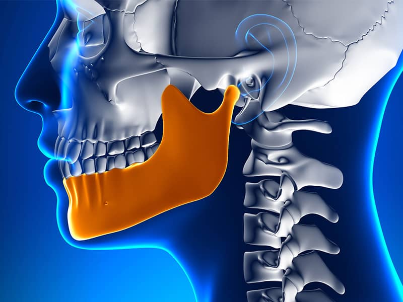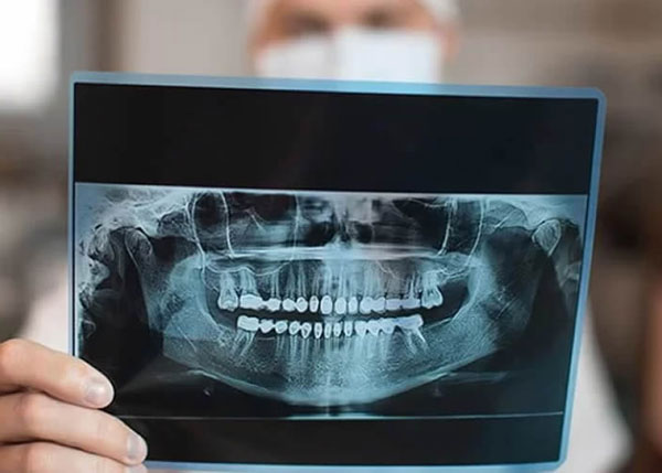
Dental tomography is an advanced imaging method that allows detailed examination of the teeth and jaw-face area. Known as cone beam computed tomography (CBCT), this technology provides three-dimensional and high-precision images. It is used in many areas such as implant planning, evaluation of jawbone, analysis of impacted teeth, and detection of pathological formations. CBCT provides essential anatomical information for accurate diagnosis and treatment planning. However, due to factors such as radiation exposure and cost, it should be preferred only in cases where it is truly necessary. This method plays a significant role in dental practices and is one of the most effective tools offered by modern technology.
[block id=”call-to-action-v2″]
| Definition | It is a radiological examination method that provides detailed three-dimensional imaging of the teeth and jaw area. |
| Areas of Use | Implant planning, evaluation of impacted teeth, diagnosis of jawbone diseases, detection of cysts and tumors, detailed examination before root canal treatment, orthodontic planning. |
| Advantages | High-resolution and detailed imaging, measurement of bone density, and accurate diagnosis with a lower radiation dose. |
| Who is it Applicable To? | Patients planned for implant treatment, individuals with abnormalities in jaw and teeth structures, patients undergoing orthodontic treatment, those with jaw joint disorders. |
| Scanning Duration | Typically lasts between 10-20 seconds. |
| Radiation Dose | Generally lower than traditional tomography, but may be slightly higher than panoramic X-rays. |
| Is Special Preparation Required? | Usually does not require special preparation. Metal accessories and appliances should be removed. |
| Side Effects | It is not recommended for pregnant women due to radiation exposure. Other than that, there are no known side effects. |
What is Dental Tomography and How Does It Work?
Dental tomography is a medical imaging method that provides detailed three-dimensional visualization of the teeth and jaw-face area. Cone beam computed tomography (CBCT) devices offer fast and precise results using a special X-ray technology:
- Forms a cone beam with X-rays
- Rotates 180-360 degrees around the patient
- Collects two-dimensional images
- Processes the images with computer software
- Creates a three-dimensional volumetric data set
With this method, sections can be taken in axial, coronal, and sagittal planes, enabling a clear examination of complex anatomical structures. It is particularly preferred in dentistry and is effective in various applications.
The most notable advantage of CBCT is its detailed three-dimensional imaging. It provides a lower radiation dose compared to traditional methods, which is important for patient safety. However, disadvantages such as low soft tissue contrast and high device costs should also be considered. Nevertheless, the benefits it provides for accurate and effective diagnosis largely compensate for these disadvantages.
In Which Situations is Dental Tomography Used?
It contributes to accurate diagnosis and treatment planning by examining the jaw and dental area in detail. The situations where dental tomography is used include:
- Implant planning and evaluation
- Detection of impacted teeth
- Analysis of complex root canals
- Orthodontic treatment planning
- Examination of the chin tissues and tumors
- Diagnosis of temporomandibular joint disorders
- Evaluation of trauma-related fractures and injuries
- Detailed imaging of the maxillary sinuses
This technology offers three-dimensional analysis capabilities that go beyond traditional two-dimensional imaging in dentistry. Thus, the location, size, and relationship of anatomical structures are determined more accurately. Especially structures vital like nerves and vessels are preserved, making the treatment processes safer.
What are the Advantages and Disadvantages of Dental Tomography?
Advantages:
- Three-dimensional imaging
- High resolution
- Accurate diagnosis
- Precise treatment planning
- Low radiation level
- Fast scanning
- Pain-free procedure
Disadvantages:
- High cost
- Difficulty in imaging soft tissues
- Need for experienced interpretation
- Motion artifacts
- Radiation exposure
What Should Patients Expect During a Dental Tomography Procedure?
Dental tomography procedures are generally quick and effortless, prioritizing patient comfort. To avoid affecting the quality of the images, all metal accessories are removed before the procedure. Patients are positioned standing, sitting, or lying down depending on the type of device. Once the correct position is assumed, the device rotates around the head to capture images. It is important not to move during the procedure in order to obtain clear images.
Things to be removed before use:
- Accessories
- Eyeglasses
- Other items containing metal
- Devices used during positioning:
- Jaw support
- Head support
The rotation around the tomography device’s head may last from 10 to 40 seconds. You are expected to remain still during the procedure to avoid movement. Modern devices minimize this duration, completing the process more quickly. No pain is felt during the scan, and discomfort is generally at a minimum level.
After the dental tomography is completed, the three-dimensional images obtained will be analyzed by your dentist or radiologist. These images play a critical role in several areas, such as examining jaw structure, detecting dental pathologies, and detailing treatment planning. After the procedure, patients can immediately return to their daily activities, as no recovery process is required.
Is There a Radiation Risk With Dental Tomography?
Dental tomography, especially using CBCT, provides detailed imaging of the teeth and jaw areas; however, this procedure carries a certain radiation risk. Due to the biological effects associated with radiation and long-term risks, it is recommended that this method be used carefully:
- Emits higher radiation than traditional 2D dental X-rays.
- Contains 5-16 times more radiation than standard dental X-rays.
- Total radiation dose is 96% lower compared to conventional CT.
- Ionizing radiation may cause damage to tissues, including cancer risk.
- The thyroid gland is one of the organs most sensitive to radiation.
Various measures should be taken to reduce radiation exposure in the use of CBCT. It is recommended to keep the field of view (FOV) small, use low mA settings, short exposure times, and narrow beam collimation. Additionally, these measures become even more critical for sensitive groups such as children and pregnant women. The risk of thyroid cancer is higher in children, and exposure to radiation during pregnancy can lead to serious consequences for both the mother and the fetus.
International standards advocate for the justification of medical procedures through benefit-risk analysis. Therefore, CBCT should only be preferred in cases where its necessity is proven. To minimize the harmful effects of radiation, dentists should perform procedures with proper planning and optimized device settings.
What are the Differences Between Dental Tomography and Panoramic Radiography?
Dental tomography (CBCT) and panoramic radiography are two different imaging methods used in dentistry for diagnosis and treatment planning. Both provide features suitable for different situations, and knowing these differences is crucial for selecting the right method.
Imaging size and type:
CBCT: Provides three-dimensional images.
Panoramic radiography: Provides two-dimensional general images.
Detail and resolution:
-
\t
- CBCT: High resolution, suitable for fine details.
- Panoramic radiography: Sufficient for general assessment.
\t
Radiation dose:
-
\t
- CBCT: Contains higher radiation, but has been reduced with new technologies.
- Panoramic radiography: Applies lower radiation.
\t
Areas of use:
-
\t
- CBCT: Implant planning, jaw joint disorders, complex cases.
- Panoramic radiography: General dental condition, impacted tooth analysis, orthodontic assessment.
\t
Distortions and errors:
-
\t
- CBCT: Metal artifacts can be an issue.
- Panoramic radiography: Distortion and magnification errors may be observed.
\t
Both methods serve different needs in dental practice. CBCT is generally preferred for complex cases requiring detailed imaging, while panoramic radiography stands out as a more common and quicker option.
How Are Dental Tomography Results Interpreted?
Accurate interpretation of dental tomography results is critical for the success of diagnosis and treatment processes. This interpretation process is carried out by specialized radiologists, and each detail is carefully analyzed:
-
\t
- In the imaging phase, the position of the teeth and root structures
- Alveolar bone density and volume
- The location of the mandibular canal, sinus cavities, and nerves
- Structures are examined, including the periodontal ligament space.
\t
\t
\t
In order to detect pathological findings, formations such as cysts and tumors, infection foci, and periapical lesions are analyzed in the tomography. When it comes to jaw and dental traumas, the distribution of fracture lines and the degree of damage are evaluated in detail. Additionally, root canals and potential extra canals are identified before endodontic treatment:
-
\t
- In orthodontic evaluation, impacted and supernumerary teeth
- The eruption directions of the teeth
- Jaw relationships and skeletal anomalies are analyzed.
\t
\t
Dental tomography facilitates the viewing of small details thanks to its high resolution. However, certain factors, such as metal restorations, can affect image quality. During the reporting process, tomography results are combined with clinical findings for treatment planning.