

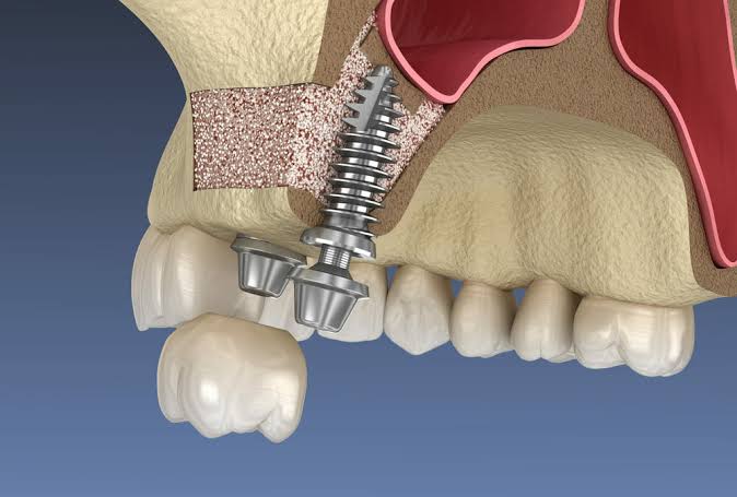
Izmir Implant Pre-sinus Lifting
Bone loss in the back regions of the upper jaw not only disrupts the chewing mechanics but also alters the physiological boundaries of the anatomical structures in that area. Particularly, maxillary sinuses—air cavities adjacent to the roots of molars in the upper jaw—gradually expand into the edentulous areas, narrowing the bone volume over time. In such cases where there is insufficient vertical bone space for the placement of titanium artificial roots, advanced surgical procedures known as “Sinus Lifting” are utilized.
What is Implant Pre-sinus Lifting and What is the Medical Purpose of Planning it?
Within the anatomy of the skull, there are maxillary sinus cavities filled with air and integrated into the respiratory system, located just inside the upper jawbone. The base of these cavities is very close to the root tips of the back molars of the upper jaw. In some individuals, the tooth roots may directly contact the sinus membrane. When an individual loses their back teeth, the bone tissue in that area begins to resorb over time due to lack of stimulation from chewing forces. At the same time, the upper sinus air cavity also sags downward, thinning the bone from within.
This anatomical narrowing makes it impossible to place a standard titanium screw into the bone. It is necessary for the tip of the screw not to fall into the void and to be surrounded by hard tissue. The sinus lifting procedure provides a medical infrastructure that supports the attraction of the body’s own bone repair cells to that area using biomaterials and the construction of real bone tissue over time to replace the artificial graft.
In Which Cases is the Sinus Lifting Operation Planned in Clinics in the İzmir Region?
Dentists make millimetric calculations based on three-dimensional dental tomography (CBCT) images when deciding on the operation. Attempting to forcefully place material into an area where bone thickness is insufficient can lead to the tearing of the sinus membrane and the escape of the material into the air cavity. It is necessary to prepare a medical ground to overcome such anatomical constraints.
| Anatomical or Pathological Condition | Effect on Bone Volume |
|---|---|
| Long-Term Tooth Loss (Atrophy) | Bone tissue that does not receive chewing stimulation is physiologically resorbed over the years, and the vertical distance decreases significantly. |
| Advanced Periodontal Diseases | Inflammation of the supporting tissues surrounding the teeth (periodontitis) creates a space structure by causing the bone to deteriorate even before tooth extraction. |
| Traumatic Tooth Extractions | Damage or loss of the thin cortical bone forming the sinus floor during difficult extractions experienced in the past. |
| Genetic / Anatomical Structure | Some individuals are structurally born with enlarged maxillary sinus cavities, which genetically limits bone thickness. |
What Are the Main Differences Between Open Sinus Lifting and Closed Sinus Lifting?
The technique to be applied is determined by the surgeon based on the clear bone distance obtained from the patient’s jaw tomography. The main principle of both methods is to separate the delicate membrane called the “Schneiderian membrane” from the bone base without damaging it and to fill the gap with biomaterial.
1. Closed Technique (Osteotomy Method): It is performed by entering through the drilling hole where the implant will be placed, without making an additional incision on the gum. The membrane is gradually pushed upwards with special hand tools (osteotomy), and a graft is placed in the small space created. Usually finished in the same session as the implant placement, the trauma is minimized.
2. Open Technique (Lateral Window Method): A flap is raised in the form of a cover from the outer tissue on the side. A small window is opened on the lateral wall of the jawbone to directly access the sinus membrane. After the membrane is lifted, bone graft material is placed in the resulting space and the area is closed and sutured with special collagen membranes (resorbable barriers).
How is the Sinus Lifting Process Managed at Avrupadent?
Interventions to the sinus area are surgical procedures that involve high precision and carry the risk of membrane tearing (perforation) when performed with traditional rotary instruments. In institutions where interdisciplinary work is conducted, “Piezosurgery” technology is included in the process for bone cutting. These devices, which operate by vibrating sound waves, stop the cutting process when they come into contact with the soft tissue of the sinus membrane, thus enhancing tissue protection to a higher level.
The management of the process is not limited to the operating table. The surgical team collaborates with prosthesis specialists during the planning phase to pre-determine the volume of the graft to be added and the length of the titanium root to be applied. The overall health profile of the patient is examined in detail, and appropriate pharmacological (medication) support is arranged to ensure infection control post-operation.
What are the Properties of Biomaterials Used as Bone Grafts?
It is a medical necessity for biomaterials to have passed international biocompatibility tests and to have been subjected to sterilization processes that will not cause tissue rejection. The particles added to the sinus cavity serve as a bridge for the body to transport calcium, phosphate, and osteoblast cells (bone-forming cells) to that area.
| Biomaterial Class | Source | Clinical Function and Reaction |
|---|---|---|
| Autogenous Graft | Obtained from the patient’s own jawbone or a different bone area. | It is the gold standard material that provides the fastest overall healing due to the presence of living cells (osteogenic). |
| Xenograft | Inorganic scaffolds that are obtained from living organisms such as cattle or pigs, stripped of all organic structures. | With a structure that can maintain its volume for a long time, it has a wide range of applications as a space-filling (osteoconductive) matrix within the sinus. |
| Alloplastic (Synthetic) | Produced chemically in laboratory settings, such as hydroxyapatite or tricalcium phosphate. | It is completely synthetic; over time, the tissue fluids are absorbed and gradually replaced by the patient’s own bone. |
What is the Schneiderian Membrane and Its Importance During Surgery?
This membrane, found in human anatomy, is a thin and quite fragile tissue. It serves as a physiological structure that filters, humidifies the respiratory air, and forms a barrier against infections in the sinus cavities. Gently mobilizing this membrane during the surgical procedure is a fundamental step reflecting the clinician’s experience in terms of the progress of the operation.
If a perforation (tear) occurs in the membrane during bone cutting or pushing procedures, the dentist applies different medical protocols based on the size of the tear. Small-sized tears are closed by patching with special collagen membranes, and the procedure continues. However, in cases of very large tears, the area is closed to eliminate the possibility of infection, and healing of the wound is awaited, after which the operation is planned for a later date.
How Long Should One Wait After Adding Bone Powder for Implant Placement?
The main element determining the improvement schedule is the titanium screw’s capacity to tightly hold onto the remaining original bone at first (primary stability). The screw should not be movable when positioned. If the doctor, based on the measurements taken through tomography, predicts that the screw’s end part can hold onto a solid bone of at least three or four millimeters, both lifting and screw placement can be done in one surgical session. This “simultaneous” approach significantly reduces the patient’s clinical period.
However, if there is a lack of existing bone structure (for example, 1-2 mm), a two-phase (two-session) procedure is preferred because the screw may rotate or sink into the sinus when it is placed. In the first phase, only bone powder is placed, and the gum is closed. The body will gradually transform these powders into a hard bone block over approximately six months. In the second surgical phase, a titanium root will be positioned within this newly formed tissue.
What is the Medical Recovery Process for Patients After Sinus Lifting Surgery?It is common to experience swelling (edema) and slight subcutaneous bruising in the operation area within the first few days following the procedure. Using prophylactic (protective) antibiotics, decongestant nasal drops, and analgesics prescribed by the physician at the specified times supports tissue comfort. Healing in the area progresses rapidly within an average of one week.
The first 48 hours after the surgical intervention is the inflammation phase in which tissues react to stress. During this period, applying cold compresses (ice) intermittently from the outside constrains bleeding by narrowing the vessels and limits the volume of swelling that may occur on the face. Sensitivities experienced as anesthesia wears off are kept at routine levels with medical medications.Medical Restrictions that the Patient Must Observe:- Avoiding Pressure: For two to three weeks following the procedure, it is prohibited to sneeze forcefully, to sneeze with the mouth closed, or to play wind instruments. An increase in air pressure within the sinuses can lead to the displacement of the implanted bone fragments.
- Sleeping Position: Keeping the head elevated above heart level (sleeping with two pillows) reduces blood flow to the head area, which helps minimize swelling.
- Fluid Intake: Drinking liquids through a straw increases the risk of bleeding due to negative pressure created inside the mouth; beverages should be sipped directly from a glass.
Can Sinus Lifting Be Performed in Older Patients or Those with Systemic Problems?
Rather than chronological age, the individual’s wound healing capacity and blood values are taken into account. Patients in the older age group can be included in this surgery if their medical history permits. However, in individuals using intravenous bisphosphonates due to uncontrolled diabetes, bleeding disorders, or osteoporosis, surgery will not be performed without consultation with a physician.In jaw surgery protocols, the patient’s medical history determines the response of bone cells. While age alone does not pose a barrier, chronic diseases associated with age shape the treatment schedule. For instance, in hypertensive patients, the choice of anesthesia may be altered due to the potential impact of procedural stress on heart rate. If there is no systemic obstruction, being older does not create a contraindication for sinus lifting.
Due to the special condition of the sinus region, patients suffering from chronic sinusitis, nasal polyps, or severe allergic rhinitis are referred to Ear, Nose, and Throat (ENT) specialists for the prior treatment of these respiratory infections. Performing surgical procedures while the sinus mucosa is already inflamed and thickened is not in accordance with medical norms, as it can lead to the added bone powder becoming infected.
How Does the Use of Tobacco Products Affect the Sinus Lifting Recovery Timeline?
Cigarettes and other tobacco products significantly reduce the amount of blood and oxygen reaching the area by narrowing the capillaries through the nicotine and toxins they contain. In the operation area where blood flow is weakened, the process of the implanted bone powders merging with living cells (vascularization) is disrupted, and the risks of wound opening or infection development increase significantly.The conversion of a biomaterial added to the body into living tissue depends on its nourishment from the surrounding blood vessels. Cigarette smoke dries out the oral cavity tissues and suppresses the activity of fibroblasts, which are necessary for cellular repair. Patients undergoing sinus lifting prior to implants in Izmir must severely restrict tobacco use at least two weeks before the surgery and throughout the bone healing phase (which can take months).
Continuing to smoke in cases of advanced surgical applications not only delays healing but also paves the way for the graft to be rejected by the body as it is expelled. This habit increases the likelihood of medical failure and is thoroughly examined in detail by dentists during the pre-operative history, with necessary warnings about physiological risks communicated to patients.
The first 48 hours after the surgical intervention is the inflammation phase in which tissues react to stress. During this period, applying cold compresses (ice) intermittently from the outside constrains bleeding by narrowing the vessels and limits the volume of swelling that may occur on the face. Sensitivities experienced as anesthesia wears off are kept at routine levels with medical medications.
- Avoiding Pressure: For two to three weeks following the procedure, it is prohibited to sneeze forcefully, to sneeze with the mouth closed, or to play wind instruments. An increase in air pressure within the sinuses can lead to the displacement of the implanted bone fragments.
- Sleeping Position: Keeping the head elevated above heart level (sleeping with two pillows) reduces blood flow to the head area, which helps minimize swelling.
- Fluid Intake: Drinking liquids through a straw increases the risk of bleeding due to negative pressure created inside the mouth; beverages should be sipped directly from a glass.
Can Sinus Lifting Be Performed in Older Patients or Those with Systemic Problems?
In jaw surgery protocols, the patient’s medical history determines the response of bone cells. While age alone does not pose a barrier, chronic diseases associated with age shape the treatment schedule. For instance, in hypertensive patients, the choice of anesthesia may be altered due to the potential impact of procedural stress on heart rate. If there is no systemic obstruction, being older does not create a contraindication for sinus lifting.
Due to the special condition of the sinus region, patients suffering from chronic sinusitis, nasal polyps, or severe allergic rhinitis are referred to Ear, Nose, and Throat (ENT) specialists for the prior treatment of these respiratory infections. Performing surgical procedures while the sinus mucosa is already inflamed and thickened is not in accordance with medical norms, as it can lead to the added bone powder becoming infected.
How Does the Use of Tobacco Products Affect the Sinus Lifting Recovery Timeline?
The conversion of a biomaterial added to the body into living tissue depends on its nourishment from the surrounding blood vessels. Cigarette smoke dries out the oral cavity tissues and suppresses the activity of fibroblasts, which are necessary for cellular repair. Patients undergoing sinus lifting prior to implants in Izmir must severely restrict tobacco use at least two weeks before the surgery and throughout the bone healing phase (which can take months).
Continuing to smoke in cases of advanced surgical applications not only delays healing but also paves the way for the graft to be rejected by the body as it is expelled. This habit increases the likelihood of medical failure and is thoroughly examined in detail by dentists during the pre-operative history, with necessary warnings about physiological risks communicated to patients.







