What Are the Aesthetic Approaches for Broken Front Teeth?Aesthetic approaches for broken front teeth are implemented through prosthetic applications supported by composite bonding, porcelain laminates, zirconium crowns, E-Max glass ceramic restorations, and root canal treatment in necessary cases, depending on the depth of the fracture, the rate of material loss, and the vitality condition of the tooth.
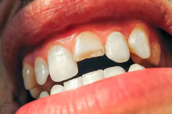
The incisors located in the anterior region of the mouth and dental health are strategic anatomical structures that directly influence an individual’s facial expression, speech mechanics, and smile symmetry. Fractures occurring in these teeth due to mechanical trauma such as impact, fall, or hard object collision can lead to both physical sensitivity and visual concerns. Repairing such structural disorders within the framework of modern medical principles is realized through the biomaterials and adhesive technologies offered by the Aesthetic Dentistry discipline. In Avrupadent clinics, these processes are addressed with a personalized medical approach by examining the patient’s anatomical structure and jaw closure dynamics.The path to be followed in the repair of fractures in front teeth varies depending on which layers of the tooth enamel, dentin, or nerve are affected. In these cases, evaluated over a wide range from superficial enamel cracks to significant tissue loss, prosthetic or conservative solutions are produced while adhering to tissue-preserving principles.
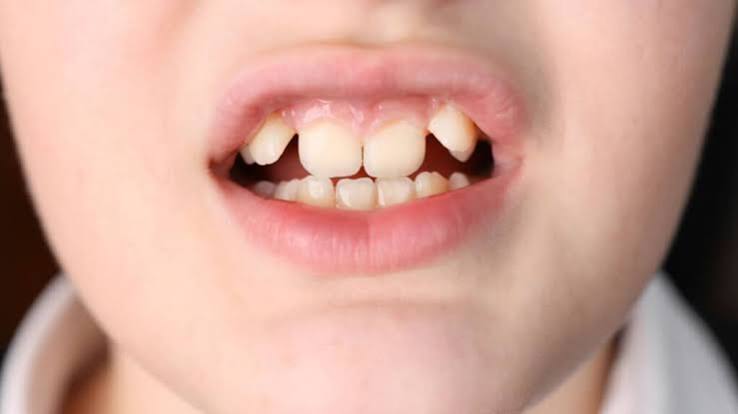
What Physical and Anatomical Reasons Lead to Front Tooth Fractures?Anterior teeth are sensitive to mechanical trauma due to their exposure to external factors; the primary physical factors that lead to these fractures include sports injuries, traffic accidents, uncontrolled impacts from hard foods, or developmental weaknesses of the tooth enamel.
The fracture line can remain within the enamel boundary, or it can progress deeper into the dentin layer or directly to the tooth nerve (pulp) chamber. This situation leads to the tooth developing sensitivity to hot and cold stimuli. In the science of anatomy, such traumatic injuries are classified based on the condition of the tooth’s root structure and the surrounding bone tissue and subject to a medical evaluation process.
The crystalline structure of tooth enamel is hard but brittle in character. When an anterior tooth suffers an impact to the front, the force spreads along the enamel prisms, causing a sudden tissue rupture. The positions of teeth in the dental arch (for example, the anterior incisors being more exposed to external impacts) are the fundamental anatomical reasons for the concentration of fracture cases in the anterior region.
How Does the Initial Examination and Clinical Evaluation Process for Anterior Fractures Proceed?
For individuals presenting to the clinic after trauma, the first step is to conduct vitality tests on the tooth and examine the root tips through radiological imaging; this evaluation is performed to determine the depth of the fracture and to detect potential cracks within the root.Radiological records guide the detection of hidden cracks in the tooth root or traumatic injuries that may occur within the bone. The dentist analyzes the size of the fractured tooth piece, the relationship of closure, and the condition of the surrounding gingival tissue. This clinical evaluation forms the basis for choosing the appropriate restorative or prosthetic method (composite bonding, laminate, or crown).
Vitality tests reveal whether the nerve and blood vessel bundle inside the tooth has been damaged or not, using electrical current or cold tests. If the nerve tissue has lost vitality, root canal treatment is planned before aesthetic restoration to prevent the risk of infection. This phase of clinical evaluation is the most critical foundational step that determines the long-term success of the treatment.
What Advantages Does Composite Bonding Method Offer for Fractured Anterior Teeth?
In small to medium-sized anterior tooth fractures, the composite bonding method applied without any enamel reduction is based on the principle of layering resin-based nano-hybrid materials onto the tooth.This method allows for the restoration of aesthetic form within a single session while preserving the tooth’s own tissue. Thanks to adhesive systems, composites chemically bond to the enamel and display optical properties that make the fracture line invisible. The different color tones and translucency degrees used during the procedure support compatibility with the natural tooth tissue of the repaired area.
Since composite bonding applications do not require a laboratory phase, they save time for the patient. This resin material, shaped directly in the chair without any loss of tooth structure, achieves a flawless surface brilliance when supported by proper polishing procedures. This method, one of the most concrete examples of a preventive approach in Aesthetic Dentistry, is an effective medical solution for mild to moderate fractures.
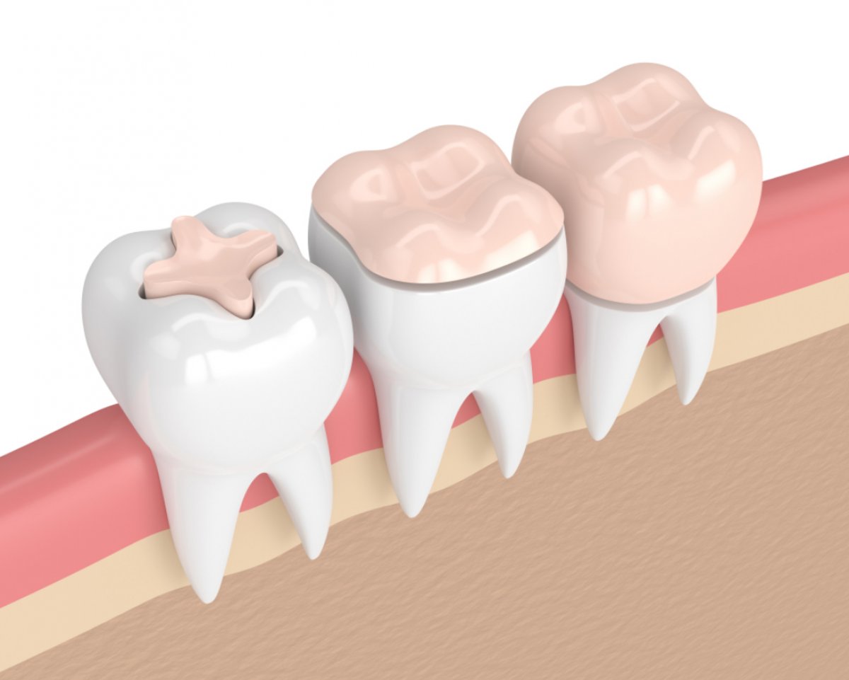
Restorative Method Application Area and Criteria Intervention to Tooth Structure Composite Bonding Small and medium-sized incisal edge fractures. No tooth reduction (Prepless). Porcelain Laminate Middle-sized cracks and surface asymmetries. Minimal enamel grinding from the front surface. Zirconium Crown Significant material loss and weakened tooth structures. Tooth is circumferentially reduced.
When Are Porcelain Laminates Planned for Traumatic Anterior Tooth Cracks?
Porcelain laminates, which consist of very thin glass-ceramic leaves covering the front surface of the tooth, are planned prosthetic alternatives for repairing middle-sized cracks and persistent discolorations.These ceramics are prepared in the laboratory by minimal grinding from only the enamel layer on the lip side of the tooth. Within the applications of Aesthetic Dentistry, the laminates, which stand out with their tissue-preserving nature, maintain the natural enamel appearance for many years due to their translucency.
Since the surface of porcelain laminates is polished and glazed, they are not affected by food pigments, and their stain resistance is high. When there are broad patterns of crack lines or enamel hypoplasias spread over a large area on the front surface of the tooth, these thin leaf ceramics exhibit effective prosthetic compatibility for both crack repair and surface renewal.
How Are Zirconium Crowns Applied to Anterior Teeth with Significant Material Loss?
In cases where a significant part of the tooth is lost due to high trauma severity, and minimal methods such as composite or laminate cannot provide structural integrity, zirconium crowns are applied.A protective infrastructure is formed around the tooth in such a way that digital measurements are taken and crowns carved from zirconium blocks are integrated into the tooth. This material provides long-term structural protection against chewing forces by offering high fracture resistance and works in harmony with the gums.
Zirconium crowns, due to their metal-free supportive structure, do not cause gray reflections at the gum line. Modern zirconium blocks, with increased light translucency, meet the aesthetic expectations of the anterior region while creating a safe protective shield mechanically by wrapping the fragile tooth roots and crown 360 degrees.
What Optical Properties Do E-Max Glass Ceramic Restorations Provide for Fractured Anterior Teeth?
Lithium disilicate-based E-Max glass ceramics provide high-level translucency (light permeability) in anterior fractures thanks to their monoblock structures devoid of a metal infrastructure.This material, which optically replicates the opalescent properties of natural tooth enamel, allows for seamless integration of the restored anterior tooth with the surrounding teeth. E-Max systems, with their high translucency and aesthetic depth, are frequently chosen as a biocompatible option for single crowns and thin veneer applications.
The interlocking crystals within E-Max glass ceramics increase the material’s fracture resistance while halting crack progression. In sensitive cases where the fractured tooth must match color and translucency with neighboring teeth in the anterior region, these glass ceramics yield successful optical results.
What Are the Prosthetic Approaches for Fractured Anterior Teeth Requiring Root Canal Treatment?
In cases where the fracture line reaches the tooth’s nerve chamber and the internal tissue is compromised, root canal treatment is primarily applied to eliminate the risk of infection.After root canal treatment, to prevent the teeth from breaking due to loss of fluid, applications of fiber posts providing internal support and then zirconium or full ceramic crown restorations are planned. This combined approach allows for long-term use while preserving the anatomical integrity of teeth that are at risk of losing their functionality.
Since the front teeth undergoing root canal treatment may change color over time, opaque ceramic substructures or zirconium crowns that mask the underlying dark root color are preferred during the prosthetic planning phase. This ensures the internal health of the tooth is protected while achieving an aesthetically pleasing appearance harmonious with other teeth when viewed from the outside.
How is Gum Health and Pink Aesthetics Maintained After Trauma?
In front tooth traumas, not only the teeth but also the surrounding gum tissue (pink aesthetics) can be affected by impact or may develop asymmetries in their levels.Before proceeding with restorative procedures, it is necessary to stabilize gum health, eliminate tissue inflammations, and, if needed, perform laser-assisted contour adjustments. A healthy gum framework is a fundamental biological element that supports the optical success of the porcelain or composite restorations applied.
If there is swelling or bleeding in the gum tissue that has been impacted, healing of these tissues should be awaited before obtaining measurements. Healthy gum boundaries (zenith points) ensure that porcelain or bonding applications to be performed in the front area exhibit a natural emergence profile, completing the aesthetic integrity.
How are Digital Measurements and CAD/CAM Systems Used in Treating Fractured Front Teeth?
In accordance with current clinical protocols, intraoral optical scanners are used to scan the fractured tooth and transfer it to the laboratory.These digital cameras, which replace physical measurements and pastes, transmit a three-dimensional point cloud map of the tooth to the computer screen within seconds. Using these data, CAD/CAM systems ensure that restorations are excavated with millimeter precision and minimize human error margins.
The transmission of digital data to the laboratory within seconds via cloud systems reduces the production time for prosthetics while also increasing the comfort of temporary restorations placed on the patient. Designing the anatomical form of a broken anterior tooth in a way that is compatible with adjacent teeth in a virtual environment is effective in achieving predictable clinical outcomes.
What Protocols Are Followed for Anterior Tooth Planning at Avrupadent Clinics?
The treatment of broken anterior teeth at Avrupadent clinics involves detailed radiological examinations, the assessment of vitality tests, creating digital design simulations in accordance with Aesthetic Dentistry standards, and protocols for selecting patient-specific materials.Every case addressed with a multidisciplinary approach is managed with a step-by-step treatment timeline aimed at prolonging the biological lifespan of the tooth and ensuring visual harmony. The physician evaluates the patient’s occlusion relationship and joint dynamics to consider the most conservative prosthetic alternative.
During the treatment process, the patient’s expectations are addressed through transparent communication, and mock-up trials of the virtual designs are conducted to test the results in advance. This meticulous clinical flow ensures both the functional health and the natural aesthetics of broken anterior teeth in the long term.
What Are the Frequently Asked Questions?
1. What should be done first when an anterior tooth is broken?
If the broken piece is available, it should be stored in a clean container and a dentist should be consulted as soon as possible to examine the vitality and root status of the tooth radiologically.
2. Can broken front teeth heal on their own?
Since the tooth enamel and dentin layer do not have the biological capacity to self-renew, it is unlikely that fractures will heal spontaneously; intervention by a physician is necessary.
3. Can composite bonding be applied to all types of front tooth fractures?
It is extremely effective for small and medium-sized fractures; however, if a large portion of the tooth is lost or the root support is weakened, porcelain crowns should be preferred.
4. Do porcelain laminates carry a risk of breaking in fractured front teeth?
Since they are chemically bonded to the enamel using special adhesive cements, the risk of breaking due to abnormal trauma, such as dislodging hard objects, is extremely low as long as such incidents are avoided.
5. Do zirconium crowns look aesthetic in the front region?
Modern translucent zirconium blocks, which increase light transmission, meet the aesthetic expectations of the anterior region and do not create metal reflections at the gum line.
6. Is local anesthesia applied during the treatment of broken front teeth?
In cases where there may be sensitivity due to the exposure of the dentin layer or in preparations where the tooth needs to be cut, local anesthesia is preferred for the comfort of the patient.
7. Do root-treated front teeth change color?
Over time, slight darkening may be observed in root-treated teeth due to loss of internal fluid, but this situation can be camouflaged using opaque bases in the prosthetic phase.
8. How is the vitality of traumatized teeth tested?
The response of the nerve tissue inside the tooth is determined using cold tests applied by the dentist or with electric pulp test devices.
9. How durable are E-Max glass ceramics in broken anterior teeth?
They possess more than sufficient compressive strength for normal functional loads in the anterior region and perform superiorly aesthetically due to their high translucency.
10. Can the broken piece be reattached to the tooth itself?
If the piece is preserved intact and clean, it can sometimes be temporarily or permanently retained to the tooth using appropriate adhesive protocols.
11. Why is digital measurement preferred in anterior tooth treatment?
It is preferred to prevent the vomiting reflex caused by physical measuring materials and to increase compatibility rates by obtaining virtual models with millimeter precision.
12. How are broken permanent anterior teeth treated in children?
Since root development in children may not be completed yet, the tooth development process is primarily supported with tissue-preserving composite bonding applications.
13. What determines the cost of broken tooth treatment?
The cost of treatment is shaped according to the method to be applied (bonding, veneer, zirconium), the quality of the materials used, and additional medical needs such as root canal treatment.
14. How is broken tooth treatment planned according to Avrupadent standards?
Radiological findings, digital scans, and the patient’s jaw closure dynamics are evaluated through a joint consultation to create a personalized medical map.
15. How should the care of anterior teeth be done after restoration?
In addition to regular brushing routines, special attention should be paid to dental floss and interproximal cleaning, and traumatic actions such as breaking hard-shelled foods with the anterior teeth should be avoided.
The incisors located in the anterior region of the mouth and dental health are strategic anatomical structures that directly influence an individual’s facial expression, speech mechanics, and smile symmetry. Fractures occurring in these teeth due to mechanical trauma such as impact, fall, or hard object collision can lead to both physical sensitivity and visual concerns. Repairing such structural disorders within the framework of modern medical principles is realized through the biomaterials and adhesive technologies offered by the Aesthetic Dentistry discipline. In Avrupadent clinics, these processes are addressed with a personalized medical approach by examining the patient’s anatomical structure and jaw closure dynamics.
The path to be followed in the repair of fractures in front teeth varies depending on which layers of the tooth enamel, dentin, or nerve are affected. In these cases, evaluated over a wide range from superficial enamel cracks to significant tissue loss, prosthetic or conservative solutions are produced while adhering to tissue-preserving principles.
What Physical and Anatomical Reasons Lead to Front Tooth Fractures?Anterior teeth are sensitive to mechanical trauma due to their exposure to external factors; the primary physical factors that lead to these fractures include sports injuries, traffic accidents, uncontrolled impacts from hard foods, or developmental weaknesses of the tooth enamel.
The fracture line can remain within the enamel boundary, or it can progress deeper into the dentin layer or directly to the tooth nerve (pulp) chamber. This situation leads to the tooth developing sensitivity to hot and cold stimuli. In the science of anatomy, such traumatic injuries are classified based on the condition of the tooth’s root structure and the surrounding bone tissue and subject to a medical evaluation process.
The crystalline structure of tooth enamel is hard but brittle in character. When an anterior tooth suffers an impact to the front, the force spreads along the enamel prisms, causing a sudden tissue rupture. The positions of teeth in the dental arch (for example, the anterior incisors being more exposed to external impacts) are the fundamental anatomical reasons for the concentration of fracture cases in the anterior region.
How Does the Initial Examination and Clinical Evaluation Process for Anterior Fractures Proceed?
Radiological records guide the detection of hidden cracks in the tooth root or traumatic injuries that may occur within the bone. The dentist analyzes the size of the fractured tooth piece, the relationship of closure, and the condition of the surrounding gingival tissue. This clinical evaluation forms the basis for choosing the appropriate restorative or prosthetic method (composite bonding, laminate, or crown).
Vitality tests reveal whether the nerve and blood vessel bundle inside the tooth has been damaged or not, using electrical current or cold tests. If the nerve tissue has lost vitality, root canal treatment is planned before aesthetic restoration to prevent the risk of infection. This phase of clinical evaluation is the most critical foundational step that determines the long-term success of the treatment.
What Advantages Does Composite Bonding Method Offer for Fractured Anterior Teeth?
This method allows for the restoration of aesthetic form within a single session while preserving the tooth’s own tissue. Thanks to adhesive systems, composites chemically bond to the enamel and display optical properties that make the fracture line invisible. The different color tones and translucency degrees used during the procedure support compatibility with the natural tooth tissue of the repaired area.
Since composite bonding applications do not require a laboratory phase, they save time for the patient. This resin material, shaped directly in the chair without any loss of tooth structure, achieves a flawless surface brilliance when supported by proper polishing procedures. This method, one of the most concrete examples of a preventive approach in Aesthetic Dentistry, is an effective medical solution for mild to moderate fractures.
| Restorative Method | Application Area and Criteria | Intervention to Tooth Structure |
|---|---|---|
| Composite Bonding | Small and medium-sized incisal edge fractures. | No tooth reduction (Prepless). |
| Porcelain Laminate | Middle-sized cracks and surface asymmetries. | Minimal enamel grinding from the front surface. |
| Zirconium Crown | Significant material loss and weakened tooth structures. | Tooth is circumferentially reduced. |
When Are Porcelain Laminates Planned for Traumatic Anterior Tooth Cracks?
These ceramics are prepared in the laboratory by minimal grinding from only the enamel layer on the lip side of the tooth. Within the applications of Aesthetic Dentistry, the laminates, which stand out with their tissue-preserving nature, maintain the natural enamel appearance for many years due to their translucency.
Since the surface of porcelain laminates is polished and glazed, they are not affected by food pigments, and their stain resistance is high. When there are broad patterns of crack lines or enamel hypoplasias spread over a large area on the front surface of the tooth, these thin leaf ceramics exhibit effective prosthetic compatibility for both crack repair and surface renewal.
How Are Zirconium Crowns Applied to Anterior Teeth with Significant Material Loss?
A protective infrastructure is formed around the tooth in such a way that digital measurements are taken and crowns carved from zirconium blocks are integrated into the tooth. This material provides long-term structural protection against chewing forces by offering high fracture resistance and works in harmony with the gums.
Zirconium crowns, due to their metal-free supportive structure, do not cause gray reflections at the gum line. Modern zirconium blocks, with increased light translucency, meet the aesthetic expectations of the anterior region while creating a safe protective shield mechanically by wrapping the fragile tooth roots and crown 360 degrees.
What Optical Properties Do E-Max Glass Ceramic Restorations Provide for Fractured Anterior Teeth?
This material, which optically replicates the opalescent properties of natural tooth enamel, allows for seamless integration of the restored anterior tooth with the surrounding teeth. E-Max systems, with their high translucency and aesthetic depth, are frequently chosen as a biocompatible option for single crowns and thin veneer applications.
The interlocking crystals within E-Max glass ceramics increase the material’s fracture resistance while halting crack progression. In sensitive cases where the fractured tooth must match color and translucency with neighboring teeth in the anterior region, these glass ceramics yield successful optical results.
What Are the Prosthetic Approaches for Fractured Anterior Teeth Requiring Root Canal Treatment?
After root canal treatment, to prevent the teeth from breaking due to loss of fluid, applications of fiber posts providing internal support and then zirconium or full ceramic crown restorations are planned. This combined approach allows for long-term use while preserving the anatomical integrity of teeth that are at risk of losing their functionality.
Since the front teeth undergoing root canal treatment may change color over time, opaque ceramic substructures or zirconium crowns that mask the underlying dark root color are preferred during the prosthetic planning phase. This ensures the internal health of the tooth is protected while achieving an aesthetically pleasing appearance harmonious with other teeth when viewed from the outside.
How is Gum Health and Pink Aesthetics Maintained After Trauma?
Before proceeding with restorative procedures, it is necessary to stabilize gum health, eliminate tissue inflammations, and, if needed, perform laser-assisted contour adjustments. A healthy gum framework is a fundamental biological element that supports the optical success of the porcelain or composite restorations applied.
If there is swelling or bleeding in the gum tissue that has been impacted, healing of these tissues should be awaited before obtaining measurements. Healthy gum boundaries (zenith points) ensure that porcelain or bonding applications to be performed in the front area exhibit a natural emergence profile, completing the aesthetic integrity.
How are Digital Measurements and CAD/CAM Systems Used in Treating Fractured Front Teeth?
These digital cameras, which replace physical measurements and pastes, transmit a three-dimensional point cloud map of the tooth to the computer screen within seconds. Using these data, CAD/CAM systems ensure that restorations are excavated with millimeter precision and minimize human error margins.
The transmission of digital data to the laboratory within seconds via cloud systems reduces the production time for prosthetics while also increasing the comfort of temporary restorations placed on the patient. Designing the anatomical form of a broken anterior tooth in a way that is compatible with adjacent teeth in a virtual environment is effective in achieving predictable clinical outcomes.
What Protocols Are Followed for Anterior Tooth Planning at Avrupadent Clinics?
Every case addressed with a multidisciplinary approach is managed with a step-by-step treatment timeline aimed at prolonging the biological lifespan of the tooth and ensuring visual harmony. The physician evaluates the patient’s occlusion relationship and joint dynamics to consider the most conservative prosthetic alternative.
During the treatment process, the patient’s expectations are addressed through transparent communication, and mock-up trials of the virtual designs are conducted to test the results in advance. This meticulous clinical flow ensures both the functional health and the natural aesthetics of broken anterior teeth in the long term.
What Are the Frequently Asked Questions?
1. What should be done first when an anterior tooth is broken?
If the broken piece is available, it should be stored in a clean container and a dentist should be consulted as soon as possible to examine the vitality and root status of the tooth radiologically.
2. Can broken front teeth heal on their own?
Since the tooth enamel and dentin layer do not have the biological capacity to self-renew, it is unlikely that fractures will heal spontaneously; intervention by a physician is necessary.
3. Can composite bonding be applied to all types of front tooth fractures?
It is extremely effective for small and medium-sized fractures; however, if a large portion of the tooth is lost or the root support is weakened, porcelain crowns should be preferred.
4. Do porcelain laminates carry a risk of breaking in fractured front teeth?
Since they are chemically bonded to the enamel using special adhesive cements, the risk of breaking due to abnormal trauma, such as dislodging hard objects, is extremely low as long as such incidents are avoided.
5. Do zirconium crowns look aesthetic in the front region?
Modern translucent zirconium blocks, which increase light transmission, meet the aesthetic expectations of the anterior region and do not create metal reflections at the gum line.
6. Is local anesthesia applied during the treatment of broken front teeth?
In cases where there may be sensitivity due to the exposure of the dentin layer or in preparations where the tooth needs to be cut, local anesthesia is preferred for the comfort of the patient.
7. Do root-treated front teeth change color?
Over time, slight darkening may be observed in root-treated teeth due to loss of internal fluid, but this situation can be camouflaged using opaque bases in the prosthetic phase.
8. How is the vitality of traumatized teeth tested?
The response of the nerve tissue inside the tooth is determined using cold tests applied by the dentist or with electric pulp test devices.
9. How durable are E-Max glass ceramics in broken anterior teeth?
They possess more than sufficient compressive strength for normal functional loads in the anterior region and perform superiorly aesthetically due to their high translucency.
10. Can the broken piece be reattached to the tooth itself?
If the piece is preserved intact and clean, it can sometimes be temporarily or permanently retained to the tooth using appropriate adhesive protocols.
11. Why is digital measurement preferred in anterior tooth treatment?
It is preferred to prevent the vomiting reflex caused by physical measuring materials and to increase compatibility rates by obtaining virtual models with millimeter precision.
12. How are broken permanent anterior teeth treated in children?
Since root development in children may not be completed yet, the tooth development process is primarily supported with tissue-preserving composite bonding applications.
13. What determines the cost of broken tooth treatment?
The cost of treatment is shaped according to the method to be applied (bonding, veneer, zirconium), the quality of the materials used, and additional medical needs such as root canal treatment.
14. How is broken tooth treatment planned according to Avrupadent standards?
Radiological findings, digital scans, and the patient’s jaw closure dynamics are evaluated through a joint consultation to create a personalized medical map.
15. How should the care of anterior teeth be done after restoration?
In addition to regular brushing routines, special attention should be paid to dental floss and interproximal cleaning, and traumatic actions such as breaking hard-shelled foods with the anterior teeth should be avoided.