What is Implantology? Who is it suitable for regarding dental treatment?
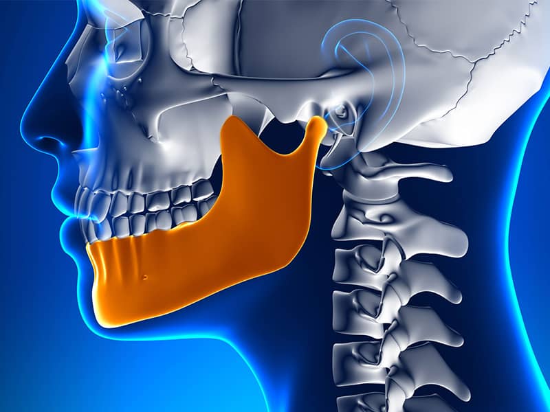
Implantology is a branch of dentistry that studies the placement of biologically compatible titanium screws into the jawbone to restore function and aesthetics where teeth have been lost. This method offers a contemporary treatment protocol for individuals who have reached adult levels of jawbone development, have an adequate general health condition, and experience tooth loss. The process is based on the biological integration of jawbone tissue and titanium material.
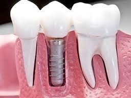
Tooth loss is a medical condition that affects many areas of individuals’ daily lives, from eating habits to speech clarity. In light of current scientific advancements in dentistry, implantology is often among the planned methods to address these deficiencies. Based on the principle of mimicking the natural tooth root, this approach serves as a foundation or infrastructure for porcelain or zirconium crowns to be added on top. The main goal of the treatment is to restore the patient’s lost chewing function and structural integrity of the oral cavity within medical standards.
What fundamental principle do Implantology Applications work on?
Implantology works on the principle of surgically placing artificial tooth roots made of titanium into the jawbone as a replacement for missing teeth. In this process, titanium screws show a biological compatibility with body tissues, demonstrating a bone integration process. This fusion, called osseointegration, aims to establish a stable foundation for the prosthetic teeth that will be added later.
The osseointegration process is when bone cells adhere to the titanium surface and create a new bone structure there. Once this biological integration is achieved, the screw in the jawbone can withstand chewing forces just like a natural tooth root. The biocompatible structure of titanium has been developed to minimize the possibility of rejection of this fusion. These implants, having undergone special treatments, possess micro indentations that facilitate bone tissue adhesion. Thus, the body recognizes this material not as a foreign substance but as a part of its own tissue, allowing the treatment process to proceed as planned.
This protocol, which involves more than just placing a screw into the jaw, requires evaluating many factors such as the anatomy of surrounding tissues, the thickness of the gum, and the position of adjacent teeth. During the planning phase, doctors use three-dimensional radiographic images to measure the angle and depth at which the implant will be placed to the millimeter. These calculations support the balanced distribution of forces that will occur during chewing across the jawbone.
Why is Implantology the Preferred Method in Cases of Tooth Loss?
Missing teeth can lead to a decrease in chewing function, speech disorders, and changes in facial symmetry. Implantology is one of the preferred current treatment protocols that allows for intervention only in the area of the gap without disturbing the healthy adjacent teeth. Additionally, the natural chewing forces transmitted to the jawbone help maintain the bone tissue in the relevant area.
In traditional bridge prostheses, the healthy teeth on either side of the gap need to be reduced (cut) to accommodate the replacement of the missing tooth. Implantology eliminates this requirement. While the anatomical structure of the healthy adjacent teeth is preserved, the gap from the missing tooth is treated as an independent unit. This approach is compatible with the protective strategies of dentistry to preserve the natural tissues inside the mouth.
After a tooth is extracted, the jawbone in that area begins to lose volume over time due to inactivity, meaning it starts to atrophy. When an implant is placed, the pressure generated during chewing is transmitted to the jawbone, just like it does under a natural tooth root. Bone cells sense this pressure, keeping the tissue in that area active. Therefore, this method provides a physiological support mechanism that allows for the preservation of jawbone volume for many years. Effectively restoring chewing force enables individuals to consume solid foods and allows the initial stage of their digestive system, which is chewing inside the mouth, to proceed within physiological norms.
Who Is Implant Treatment Suitable For?
Implant treatment can be planned for individuals who have completed the bone development process (generally aged 18 and older), have sufficient density and volume in the jawbone, or for whom this volume can be achieved with bone graft (powder), and whose general systemic health status is suitable for surgical operation. Patients who pay attention to oral hygiene and comply with regular doctor check-ups are among suitable candidates.
Age is not a criterion that forms an upper limit in this treatment. The lower limit corresponds to the period when jawbone growth is completed. An implant placed while bone development continues in young individuals may remain lower than or in a different position from other teeth over the years due to ongoing jaw growth. Therefore, it is advised to examine the bone development of individuals under 18 radiologically, and if growth is incomplete, to postpone the process.
The individual must have sufficient bone thickness and height to surround the implant, which is crucial for the long-term stability of the application. Candidates with insufficient bone volume are not deprived of treatment options; advanced surgical techniques such as sinus lift or the addition of bone powder (graft) are primarily applied to prepare the medical ground for these patients. Any individual whose systemic health is adequate and whose wound healing capacity is within normal limits can be included in this process as a result of clinical and radiological evaluations conducted by their physician.
What Health Conditions Can Implantology Applications Prevent?
Uncontrolled diabetes (sugar disease), having received radiation therapy from the head and neck area recently, and the active use of bisphosphonate group medications that affect bone metabolism are fundamental health conditions that may hinder implantology applications. In these cases, special consultations with a physician are required because wound healing and bone fusion may be negatively impacted.
When blood sugar levels are irregular in diabetic patients, the body’s wound healing mechanisms slow down, and the risk of infection increases. If a patient with type 2 diabetes has an HbA1c value within the reference ranges set by the physician, implantation procedures can be planned according to standard protocols. However, in cases where values are very high and inconsistent, it is expected that blood sugar will be brought under control, initially in consultation with an endocrinologist.
Individuals using bisphosphonate-type medications for the treatment of osteoporosis (bone loss) have a risk of necrosis (bone death) in the jawbone. Since these medications disrupt the bone cycle, the healing capacity of the bone may be impaired after the surgical procedure of implants. Similarly, in individuals who receive intensive radiation therapy to the jaw area due to cancer treatment, since blood flow in the region is reduced, the integration of titanium screws into the bone becomes challenging. In autoimmune diseases such as rheumatoid arthritis that suppress the immune system, the management of the process is shaped with the joint approval of medical professionals based on the individual’s specific health condition.
What Stages Does the Implantology Process Involve?
Implantology process consists of four main stages: First, a detailed clinical examination and three-dimensional tomography are conducted to determine the diagnosis. In the second stage, the titanium root is placed through a surgical operation under local anesthesia. In the third stage, several months of bone healing are expected for osseointegration. Finally, in the last stage, the permanent prosthetic teeth are fixed onto the healed implant.
1. Clinical Examination and Planning: The individual’s oral cavity is examined radiologically. The density of the jawbone, the position of nerve channels, and the distance of sinus cavities are measured millimetrically through a panoramic X-ray or three-dimensional dental tomography (CBCT). Based on the digital data obtained, the physician decides which area will be treated, the length, and diameter of the material to be placed.
2. Surgical Phase: Following planning, the area where the procedure will take place is numbed with local anesthesia. The gum is opened to reach the jawbone. Using medical instruments, an area suitable for the size of the implant is created within the bone, and the titanium screw is placed in this area. Then, the gum is closed again and secured with stitches. This process usually takes between 20 to 30 minutes for a single tooth.
3. Healing and Integration (Osseointegration): To achieve complete biological compatibility between the screw and the jawbone, an average of 2-3 months is awaited for the lower jaw and about 3-4 months for the upper jaw. This waiting period may vary depending on the bone structure and any additional surgical procedures applied (such as bone grafts, etc.).
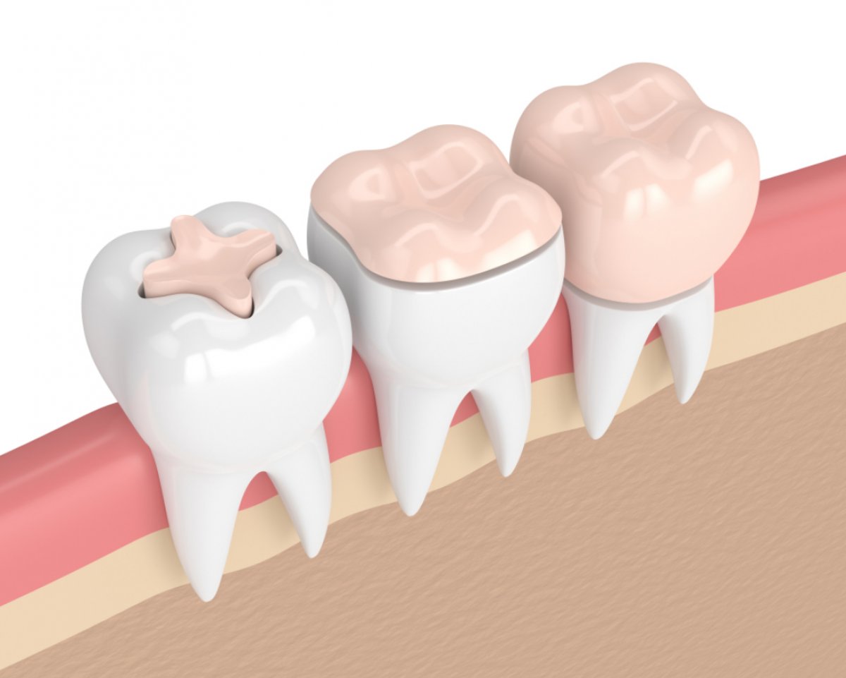
4. Prosthesis Staging: After the healing process is completed, a medical piece called “healing cap” is placed on top of the implant, and the shaping of the gum tissue is ensured. A few days later, measurements are taken inside the mouth using digital or traditional methods. The process is completed by screwing or securing a zirconium or porcelain crown (veneer), prepared in the laboratory, onto the titanium root with dental adhesives.
How is Implantology Advanced in Case of Bone Insufficiency?
If the jawbone has deteriorated and lost sufficient volume due to prolonged tooth loss or gum diseases, direct implant placement is not possible. In this case, additional surgical procedures called “bone grafting” (application of bone powder) or “sinus lifting” (lifting of the sinus) on the upper jaw are applied to increase the bone volume in the area and prepare a suitable foundation for the implant.
When many years have passed over the missing teeth, vertical or horizontal resorption occurs in the jawbone. It is essential to have bone tissue of a certain thickness around the implant for long-term functionality. A screw placed in bone that has thinned out or reduced in height may become visible from the outside over time or lose its stability underneath. To manage this situation, bone grafts sourced from synthetics or the individual’s own body are used to support the relevant area. A biological maturation period ranging from 3 to 6 months is generally expected for the grafted area to convert to its own bone tissue.
In the upper jaw’s background regions, there are anatomical air cavities known as “maxillary sinuses.” After tooth loss, these cavities can sag downwards, narrowing the bone volume. In the procedure called “Sinus Lifting,” the membrane forming the base of this air cavity is gently lifted upwards, and bone graft material is placed into the created space. In some cases, the implant placement can be performed in the same session as the bone graft application, while in cases where the amount of bone is very insufficient, it may be necessary to wait for the complete integration of the bone graft before proceeding with implant placement a few months later.
What Are the Main Differences Between Implantology and Traditional Prosthetic Approaches?
Implantology aims to restore the patient’s oral functions, but structurally differs from traditional prostheses (bridges or removable dentures). Implantology directly receives support from the jawbone, while traditional prostheses gain support from existing healthy teeth or gum tissue. This affects chewing capacity, the preservation of bone structure, and the treatment timeline.
The approach to be chosen in treatment planning is assessed together with the physician, in line with the individual’s clinical condition and physiological structure. The table below presents the clinical characteristics of these two different approaches for comparison:
| Assessment Criteria | Implantology Applications | Traditional Prostheses (Bridge/Denture) |
|---|---|---|
| Effect on the Jawbone | Transmits chewing force directly to the bone, maintaining physiological stimulation. | Since the functional loading to the bone was not done correctly, it may continue to melt over time in the edentulous area. |
| Relation with Neighboring Teeth | No cutting or grinding procedures should be performed on the healthy teeth adjacent to the gap. | To receive support in bridge systems, the healthy teeth next to it need to be reduced in size. |
| Comfort of Use and Stability | The titanium root provides a stability similar to a natural tooth since it integrates completely with the bone. | In removable dentures (palates), slight movements may occur during speaking or chewing. |
| Care and Cleaning | Care is performed with brushing, dental floss, and interdental brushes just like natural teeth, and they cannot be removed. | It is expected that removable dentures are cleaned daily by being taken out of the mouth. The underside of bridges should be cleaned with special threads. |
| Treatment Duration | Since bone union is expected, the process may take several months (if graft procedures are included, the duration is extended). | Measurements and laboratory analyses can be completed within a few weeks, allowing treatment to be finished in a shorter time. |
How Should Daily Life and Care Routine Be After Implantology?
Care in the oral cavity after implantology is a clinical necessity for the long-term function of the treatment. Individuals should brush their teeth at least twice a day and clean the plaque accumulation around the crowns using dental floss and, particularly, interdental brushes. Regular dental check-ups every six months should be an indispensable part of the routine.
Although titanium material does not pose a risk of corrosion, the gum tissue surrounding the implant is still susceptible to inflammation, just like in natural teeth. This condition, known as “peri-implantitis,” occurs when bacteria accumulate in the gum pocket due to inadequate oral hygiene and reach the jawbone, causing an infection. As the infection progresses, there can be losses in the jawbone, which may affect the retention of the titanium screw.
It is recommended to prefer toothpaste with low abrasive content in daily care routines and to use oral irrigation devices (water flosser) to remove food residues from the gum line. During regular check-ups, the dentist examines the bone level by taking X-rays when necessary, in addition to the clinical examination. Thus, any potential plaque accumulation or minor infection can be detected early, allowing for necessary interventions (such as professional dental cleaning, etc.).
Why is Titanium Material Used in Implantology?
Titanium is a metal found in nature and shows a high degree of biological compatibility (biocompatibility) with human body tissues. Its non-toxic and non-allergenic nature, along with its light weight and physical resistance to chewing pressures, makes it accepted as a fundamental structural material to be used in the field of implantology.
Titanium, which is used not only in dentistry but also in orthopedic joint prostheses and neurosurgery, is one of the metals with a low probability of being recognized and rejected as a foreign body by the body’s immune system. Various alloyed versions of pure titanium offer mechanical durability that can withstand the crushing and chewing forces generated by the back teeth without deforming.
In recent years, implants made from “Zirconia,” a type of biocompatible ceramic, have also been developed as an alternative to titanium. These white-root forms can be considered for patients with very thin gum tissue to prevent gray reflection or in cases where metal sensitivity is suspected. However, due to the extensive literature support and compatibility of titanium-based systems with bone, such systems continue to be planned in current dentistry clinics.
How Do Clinical Procedures Work in the Implantology Process?
Implantology processes are shaped within the framework of standard medical protocols, beginning with clinical patient acceptance. The current oral health status of individuals is examined in detail through radiological investigations to create a personalized treatment plan. A systemic health assessment is made before surgical intervention, and medical procedures proceed according to scientific principles throughout the process. The dentists at Avrupadent also conduct the processes based on these general medical standards.
The first phase of the treatment process involves the clinical evaluation that is realized. Utilizing three-dimensional dental tomography technologies, the anatomical structure of the jawbone, bone density, the location of nerve tissues, and the openings of adjacent tooth roots are examined. This preliminary analysis process is necessary to predict potential anatomical situations that may arise in the future and to plan for the operation to be executed in accordance with medical standards.
Surgical sterilization protocols are followed. To keep the patient’s condition under control during the hospital procedure, the area to be intervened is numbed with a local anesthetic. During the operation, not only the current bone structure but also the openings that will be suitable for the form of the prosthesis to be made in the future are calculated. Throughout the healing period, the patient is monitored, and once osseointegration is achieved, a prosthetic superstructure is added with medically appropriate materials (zirconium, porcelain) to complete the functional process.
How Does Smoking Affect the Implantology Process?
Smoking slows down the blood circulation in the mouth due to the chemicals it contains and the heat it generates. The decrease in blood flow negatively affects wound healing after surgical operations and the integration of titanium with bone. Because this situation can increase the risk of infection, it is recommended by doctors to refrain from tobacco products before and after the procedure.
Nicotine has a constrictive effect on blood vessels (vasoconstrictor). A surgical area needs oxygen, nutrients, and defense cells to heal; these are transported to the area via blood. In individuals who smoke, the amount of blood reaching the gum tissue and jawbone decreases, slowing down the body’s healing response. This physiological condition not only prolongs the integration process but also prepares the ground for the opening of stitch areas and bacterial leakage.
In long-term use, tobacco products increase the risk of developing gum diseases (periodontitis). Even years after the implant is placed, tobacco consumption can cause damage to the surrounding gum tissues (peri-implantitis). If bone resorption occurs as a result of this disease, the stability of the fixed titanium root may be compromised. Therefore, when the decision for the operation is made, it is supportive from the biological aspect of the process for individuals to reduce their tobacco habits or to quit under medical supervision.
How is Implantology Planned in Cases of Multiple Tooth Loss?
In cases of multiple missing teeth, it is not mandatory to place a separate titanium screw for each missing tooth. In adjacent gaps, bridge systems can be planned where there are implant roots at both ends and body teeth in the middle. In cases of complete tooth loss, concepts like All-on-Four may also be evaluated depending on the jaw structure.
For example, if there is a gap of three adjacent teeth in the posterior region, one implant is placed at each end of the gap. Between these two structures, a porcelain or zirconium bridge representing the three teeth is fixed. This approach overcomes anatomical constraints, preventing unnecessary surgical procedures, and mechanically distributes the burden on the jawbone. This method can be applied in any region with adequate volume, suitable for the anatomy of the jawbone.
In cases where all teeth are missing or a decision has been made to extract remaining teeth, specific medical approaches referred to as “All-on-4” or “All-on-6” can be planned. In these concepts, special implants are placed in the lower jaw or upper jaw with certain openings (usually 4 or 6). If there are anatomical barriers such as sinus cavities or nerve canals in the back parts of the jaws, a placement is performed with openings avoiding these areas, supporting a fixed prosthesis that covers the entire jaw. In suitable cases, provisional prosthetics can be placed on these stabilized screws on the same day or within a few days after the surgical operation, after which the final prosthesis stage is proceeded.
Frequently Asked Questions (FAQ) About Implantology
1. How long does the implantology procedure take?
The duration of the surgical operation varies depending on the number of screws to be placed and the condition of the bone. The placement of a single titanium root in the jaw generally takes between 20 to 30 minutes. However, this time may extend if multiple extractions are necessary or if bone grafting is required during the same session.
2. Is there an age limit for implantology?
There is no upper age limit as long as the general systemic health condition is suitable; it can be planned for individuals in older age as long as the health permits. However, the lower age limit is generally considered to be around 18 years, when bone growth and development are completed. Whether growth is still ongoing is determined by physicians through radiological data.
3. Is local anesthesia used during the treatment?
Yes, during the procedures, the relevant area is numbed with local anesthesia under the supervision of the dentist. Just like in a standard dental filling or tooth extraction, the nerve transmission in the area is blocked to ensure that the surgical intervention is completed in a controlled manner and can be managed comfortably from the patient’s perspective.
4. Can people with titanium allergies get implants?
The incidence of allergic reactions to pure titanium or titanium alloys used in dentistry is reported to be quite low in the literature. However, if there is known or suspected metal sensitivity, specific allergy tests are conducted. In such exceptional cases, doctors may consider implant alternatives made from zirconia (ceramic) material.
5. Can diabetic patients benefit from implantology?
Diabetic patients whose blood sugar levels are regularly controlled with medication, diet, or insulin can benefit from this treatment. Before the procedure, laboratory values such as HbA1c are requested, and consultation with medical professionals is facilitated. In uncontrolled diabetes, as the risk of infection increases, wound healing may be slower than expected; therefore, it is advisable to stabilize the values beforehand.
6. Do implant-supported prosthetics look like natural teeth?
The zirconia and porcelain materials used in today’s dental laboratory technologies exhibit properties similar to the structure of natural tooth enamel in terms of light transmittance. When the gum shaping is appropriately done and the correct color matching is ensured, the prosthetic structure visually integrates well with the other natural teeth in the mouth.
7. How much does bone grafting extend the implantology process?
The duration of biological integration varies depending on the amount of graft (bone powder) used and the structure of the area treated. If only minor support is provided, treatment can proceed immediately; however, if a significant volume is intended to be created, it may take an additional waiting period of 3 to 6 months for the graft to transform into the patient’s own bone structure.
8. Are temporary teeth installed until the implantology process is completed?
Especially for anterior tooth deficiencies, temporary prostheses can be planned to help patients maintain their aesthetics and speech function. These temporary teeth can be fitted in a movable form that does not burden the titanium root and attaches to adjacent teeth, or they can be adjusted by the physician within clear plates.
9. Can implantology be performed on the same day as tooth extraction?
This method, called “immediate implantation”, can be planned if the patient’s bone thickness is sufficient and there is no acute infection around the extracted tooth. Immediately after carefully removing the tooth root, a screw that is suitable in diameter can be placed in the same space. This can support the process by reducing the number of surgical sessions.
10. How should nutrition be after implantology?
In the first few days after the operation, it is recommended to consume soft, non-particle, and liquid foods (soup, puree, yogurt) to prevent damage to the stitches and protect the wound area. It is advisable to avoid very hot or cold beverages and hard, crunchy foods. Once the healing process is complete and permanent prosthetics are placed, individuals can return to their regular dietary habits.
11. Are the teeth on which implantology is performed at risk of decay?
Artificial dental materials such as titanium, zirconium, or porcelain have no risk of decay (cavitation). However, plaque can accumulate around them, and this bacterial layer can inflame the gums, leading to bone loss. Therefore, regular brushing and flossing routines are necessary to protect the biological foundation, even if there is no decay.
12. Does osteoporosis (bone loss) hinder implantology?
Osteoporosis alone may not be a barrier. The decrease in overall bone density in the body does not always directly correlate with the condition of the jawbone. However, if medical drugs containing bisphosphonates are used for osteoporosis treatment, the healing potential of the jawbone may be affected, so the process should be managed with approval from orthopedics and endocrinology specialists.
13. What X-rays are taken during an implantology examination?
In the initial examination, a panoramic X-ray is usually taken that shows all the teeth and the jaw in two dimensions. However, when it comes to the surgical planning stage, Dental Volumetric Tomography (CBCT) is used to evaluate the bone’s millimetric thickness, sinus depth, and nerve canals in three dimensions (3D).
14. Can individuals who grind their teeth at night (bruxism) get implants?
In individuals with bruxism, uncontrolled excessive forces reflect on the teeth and jaw during sleep. This situation can cause the titanium screw to move before it fuses completely with the bone or to cause cracks in the porcelain above it afterward. In these patients, medical relaxants applied to the chewing muscles after the procedure and the use of a night guard (protective plate) will help balance the process.
15. What are the clinical standards in planning implantology treatment?
Clinical standards include examining the individual’s medical history, 3D radiological planning, and the application of sterilization procedures according to standard medical rules. By conducting special analyses on the jaw anatomies of patients, all steps from prosthesis design to surgical operation are carried out, adhering to general medical principles. The approach applied in institutions like Avrupadent is also based on these scientific foundations.