

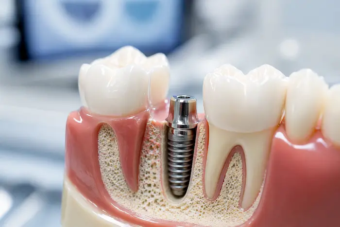
When is an implant done after sinus lifting?
The loss of the back molars in the upper jaw (maxilla) region reduces the vertical height of the jawbone over time, paving the way for the downward sagging of the maxillary sinus air cavities present in the skull anatomy (pneumatization). This two-way anatomical narrowing eliminates the necessary physiological minimum bone distance required for the placement of titanium artificial roots in place of the lost teeth. To overcome this structural limitation, advanced tissue engineering procedures, referred to as “Sinus Lifting” (Sinus Elevation), are applied in the practice of oral and maxillofacial surgery.
When is an implant done after sinus lifting and how is the healing schedule determined?
Bone augmentation procedures applied in jaw surgery are based on manipulating the body’s own self-repair (regeneration) mechanism. The particles placed in the cavity created by elevating the sinus membrane are initially inert mineral matrices. For the body to incorporate these minerals into its skeletal system, a biochemical activity that lasts for months must be initiated.
The main reason for extending the improvement timeline over a broad range (4-8 months) is the patient’s basal (existing) bone thickness. The body nourishes the added graft materials through capillaries that extend inward from living bone walls. The larger the bone defect, the longer it takes for the vessels to encompass that broad area and for bone-forming cells (osteoblasts) to reach the region. After it is objectively observed during radiological controls that the new tissue has reached the strength to support titanium material, the second surgical phase (screw placement) is conducted.
In Which Anatomical Conditions Can Simultaneous (Single-Stage) Titanium Root Application Be Performed?If the existing vertical bone thickness between the sinus cavity and the crest (bone ridge) in the patient’s jawbone is over 4-5 millimeters, the physician can perform the placement of the titanium material during the same surgical session with a sinus lifting procedure; thus, the patient’s total clinical waiting time is minimized, advancing the process with a single operation.
This approach in dental medicine literature is classified as “Simultaneous Surgery” or “Single-Stage Procedure.” For a titanium screw to complete the process of osseointegration within its natural limits, it must be tightly compressed with a high torque force upon placement in the bone (primary stability). If the patient has 4-5 millimeters of their own original bone, the lower part of the screw will hold firmly to this living bone, while the upper part will remain within the bone powders added to the sinus.In this anatomical scenario, the screw will remain fixed in place thanks to the physical support it receives from the lower part. During the biological waiting phase (averaging six months), both the living bone in the lower part cellularly integrates with the screw, and the bone powders in the upper part solidify, encasing the apex of the screw. The single-stage protocol is a tissue-friendly clinical approach that spares the patient from the stress of undergoing surgery twice (two tissue openings) within a six-month period.
What Are the Physiological Factors Determining Waiting Time in Two-Stage Surgical Approach?
In cases where the existing vertical bone thickness drops to as thin as 1-3 millimeters, a titanium screw may not achieve initial retention, making a two-stage surgery necessary. In the first session, only sinus lifting and grafting (bone powder) are performed, an approximate six-month waiting period is anticipated for the created volume to harden, and material is placed on this newly stabilized bone in the second session.If the jawbone thickness is 1 or 2 millimeters, when the surgeon attempts to place a screw in that area, the screw cannot anchor in the bone and tends to rotate around itself or show a tendency to drop into the sinus cavity. Bone tissue cannot form around a titanium material exposed to micro-mobility (vibration); the body rejects the material by encasing that area with fibrous connective tissue.
Medical Differences Between Single and Two-Stage Surgery Clinical Parameters Simultaneous (Single Stage) Procedure Stepwise (Two-Stage) Procedure Anatomical Necessity Minimum 4-5 mm existing vertical basal bone. 1-3 mm inadequate basal bone height. Total Biological Waiting Average 6 Months (Graft and material heal simultaneously). Average 9-12 Months (6 months for graft hardening + 3 months for screw integration). Number of Surgical Sessions 1 Surgical Session. 2 Separate Surgical Sessions.
How Are Sinus Lifting and Titanium Root Processes Planned Within Avrupadent in the İzmir Region?
Processes at Avrupadent, one of the equipped institutions in Izmir, involve creating a millimeter map of jaw anatomy through three-dimensional dental tomography (CBCT). Multidisciplinary physician boards analyze the volume of the sinus cavity and the remaining bone profile in a virtual environment, making simultaneous or staged surgical decisions based on objective radiological data.In the planning of advanced surgical interventions, there is no place for assumptions or two-dimensional incomplete data. While a standard panoramic X-ray roughly shows the size of the sinus sagging, it does not reflect the width of the bone or a possible pathology inside the sinus (such as the presence of a cyst or polyp). Three-dimensional volumetric scans allow for viewing the jaw and skull cavities in sections.
In institutional organizations, while the surgeon is planning, they virtually elevate the sinus membrane on the software and pre-calculate how many cubic centimeters (cc) of bone dust (graft) will be needed for that cavity. By virtually placing the titanium screw, it is clarified at the table before the operation whether sufficient compression (primary stability) can be achieved in the first session. This analytical approach minimizes the surgical time of the patient in the chair while limiting the chances of complications.
How Are Biomaterials (Grafts) Added to the Sinus Cavity Converted into Living Tissue?
Biomaterials placed beneath the sinus membrane serve as a structural cellular scaffold. The body grows new capillaries between these particles through the process of “angiogenesis”; over time, repair cells that reach the area via blood assimilate these inorganic particles into living, dense human bone that belongs to its own system (osteogenesis).The fundamental philosophy of tissue engineering is to support the body’s own repair dynamics. The bone powders used in jaw surgery are obtained from either inorganic skeletons from animal or bovine sources (xenografts), human-derived native tissues (allografts), or synthetically produced calcium phosphate compounds (alloplastic). These materials have been sterilized and are fully free of their proteins, featuring porous structures.
Stages of Bone Healing (Consolidation) Biology:- Inflammation (1-2 Weeks): Following surgical trauma, the area fills with blood. The resulting blood clot surrounds the bone powders, releasing growth factors for repair cells.
- Vascularization (2nd Week – 3rd Month): Micro-capillary vessels emerging from neighboring living bone walls create tunnels into the graft mass. The region is supplied with oxygen.
- Remodeling / Restructuring (3rd Month – 8th Month): Osteoclasts (resorptive cells) gradually dissolve inorganic powders, while osteoblasts (constructive cells) replace them with hard human bone (matrix). The area achieves mechanical strength.
What Causes Bone Resorption (Atrophy) and Sinus Drooping in the Back Area of the Upper Jaw?
Following the extraction of teeth, the bone in the back area of the upper jaw is physiologically resorbed (atrophy) due to the absence of mechanical stimulation. Simultaneously, the air pressure within the maxillary sinus above expands towards the underlying bone, causing the sinus membrane to sag downwards (pneumatization) and leading to thinning of the bone internally.Maxillary sinuses are air spaces that filter and warm the inhaled air, balancing the weight of the skull. The base of these spaces follows a course that is quite close to the roots of the molar teeth. As long as the tooth roots are in that area, the downward movement of the sinus membrane is mechanically restricted. The roots, which serve as a natural barrier, encroach on the system’s boundaries.
When one or more molar teeth are lost, not only does the bone outside deteriorate; the internal balance of the system is also disrupted. According to the biological rule “unused tissue atrophies,” as bone volume is lost, the air pressure created within the sinus expands toward the base, which does not face resistance. Over time, the vertical distance between the gum and the sinus membrane can decrease by one or two millimeters. The sinus lifting operation is planned to surgically elevate these damaged physiological boundaries back to their former anatomical level.
What Are the Differences in Healing Time Between Open Sinus Lifting and Closed Sinus Lifting?
The closed technique is less traumatic and has a shorter healing period as it involves creating a small socket from the ridge crest when the bone height is relatively sufficient. In contrast, the open technique requires lifting the gum from the cheek area to create a wide window in the bone and uses more grafts, resulting in a longer biological waiting period.The technique to be used is determined by the physician based on the existing bone thickness in the patient’s tomography images. Although the medical purpose of both procedures is the same (to elevate the membrane and place bone powder in between), the application methods differ the extent of the surgical wound.
Comparative Medical Analysis of Surgical Techniques Procedure Clinical Process Conditions of Use and Healing Timeline Closed Sinus Lifting (Osteotomy Technique) The gum cannot be opened. The area where the screw will enter is carefully broken open with specialized tools, and the membrane is pushed upwards. This is planned for cases where the bone is between 4-6 mm. Since no extra surgical wounds are created, local edema is at a minimal level. Open Sinus Lifting (Lateral Window Technique) The gum is lifted from the cheek side (flap), and a window is opened on the side of the bone to elevate the membrane directly underneath it. This procedure is mandatory in cases where the bone remains extremely thin, like 1-4 mm. Because of the intense tissue manipulation involved, 6-8 months of cellular consolidation is expected.
What Medical Rules Must Patients Pay Attention to During the Biological Waiting Period (Consolidation Phase)?
After the operation and during the waiting phase, it is essential for patients to avoid actions that can create pressure inside the mouth or nose (such as severe sneezing, closed-mouth coughing, and using straws). These types of mechanical pressures can lead to tears in the sinus membrane or displacement of the added bone powders, making it a crucial medical rule to avoid traumatizing the wound area.Interventions in the sinus cavity require careful working over the sensitive barrier between the oral cavity and the respiratory system. The sinus membrane, referred to as the “Schneiderian membrane,” is quite thin and flexible. The inorganic dust that settles beneath this membrane is extremely fragile against external factors until it coagulates with blood and hardens over months.
Especially within the first three or four weeks following the operation, a sudden change in pressure in the upper respiratory tract can create strong pressure from inside out on the sinus membrane that has not yet stabilized (for example, during airplane travel, balloon blowing, doing diving sports, or forcefully sneezing). If the membrane is torn, the added bone graft materials can spill into the sinus cavity (respiratory area), which may cause inflammation of the graft material (sinusitis). Adhering to the physician’s recommendations for the healthy maintenance of the healing period is a biological necessity.
Is Immediate Loading Applicable in Sinus Lifting Cases?
In titanium roots placed simultaneously with sinus lifting, since a large part of bone stability is supported by graft materials, immediate loading procedures (placing prosthetics on the same day) are not medically applicable. An early chewing force exerted on the material can lead to micro-movements, causing the still soft bone powders to disperse and disturbing biological healing.The early loading protocol is a method that can be planned in cases where the bone is very hard and dense, particularly in the upper jaw region (or lower jaw), with titanium material being tightly fixed to the bone under high mechanical torque. However, in the posterior area of the upper jaw, both the structure of the bone is spongy, and a significant portion of that bone is composed of added dead bone powders.
In a sinus lift procedure, a temporary tooth is placed immediately in the area where a screw is inserted during the same session. If the patient chews in that area, it may crush or disrupt the bone debris around the screw. Since the repair cells will not be able to encapsulate a vibrational material, that area will heal with flexible connective tissue instead of bone (fibrointegration). This situation completely eliminates the medical stability of the procedure, making “delayed loading” (creating a prosthesis months later) a universal medical principle to protect tissue health in sinus surgeries.
What Are the Effects of Systemic Diseases on Healing and Waiting Period After Sinus Lift?
In individuals with diabetes or circulatory system disorders, the formation of capillaries (microvascularization) will progress more slowly, which may prolong the period for the added bone debris to turn into live tissue. When the blood values stabilize with consultations from relevant branch physicians, the procedure can be performed. In such cases, the biological waiting period is extended for a few months to support cellular repair.The repair dynamics of the human skeleton work in direct proportion to the overall performance of the circulatory and connective tissue systems. For the sinus lift operation to be successful, new blood vessels (angiogenesis) must reach the area quickly. In uncontrolled Type 2 diabetes patients, high circulating glucose in the blood damages cell walls, slowing down oxygen transport and collagen synthesis.
If the injured tissues cannot receive enough oxygen, the added biomaterials cannot be assimilated by the body, and the potential for an inflammatory (necrotic) reaction in that area increases. To manage this clinical picture, doctors request patients in the risk group to have their pre-operative HbA1c (blood sugar for the last three months) values drawn within reference ranges. After the surgical procedure, if a normal patient is expected to heal in six months, for a patient with diabetes or a history of osteoporosis, this time may be extended to eight or nine months, allowing time suitable for the physiological pace of the body.
How Does Tobacco Consumption Affect Sinus Membrane Repair and Biomaterial Integration?
The carbon monoxide and toxic agents in tobacco smoke limit the oxygen transported to the surgical site by constricting the blood vessels within the mucosa. The inability of the region to receive blood slows down the repair of the sinus membrane and restricts the integration of the added bone powders with living cells. To ensure that wound healing is not interrupted, it is a medical necessity to limit these smoking habits.The sinus membrane is an extremely rich structure in terms of capillaries, which warm and humidify the respiratory air. During surgery, when this membrane is stripped from the bony base, it suffers micro-trauma. For the membrane to recover and nourish the inorganic powders placed beneath it, it requires a continuous blood flow.
When smoking, nicotine that enters the bloodstream creates a vasoconstrictive effect, blocking the defense cells and nutrients that reach this area. In an oxygen-deprived environment, the body’s wound healing cells (fibroblasts) cannot organize. The most aggressive factor that increases the risk of infection or failure in advanced surgical procedures in dentistry (especially graft applications) is the use of tobacco. Restricting this consumption before the operational schedule and during the months-long healing process is essential for the continuity of medical protocols.
How is the End of the Waiting Period for Radiological Imaging (3D Tomography) Decided?Three-dimensional dental tomographies taken after the completion of the cellular donation months objectively map whether the added bone powders (grafts) achieve a radiopaque (solidified and dense) appearance. The bone density is measured using the Hounsfield Unit (HU) value, confirming with scientific data that the area is ready for the second surgical phase (placing prosthetics or materials).
The conclusion of the waiting schedule in advanced surgical processes cannot be based solely on an estimated timeline like “six months have passed, so let’s proceed with the prosthetics.” Human metabolism is unique to each individual; while one patient’s bone may harden like stone in five months, another’s ossification may take eight months. At this point, clinical diagnosis is based on objective grounds provided by technological radiological data.
This approach in dental medicine literature is classified as “Simultaneous Surgery” or “Single-Stage Procedure.” For a titanium screw to complete the process of osseointegration within its natural limits, it must be tightly compressed with a high torque force upon placement in the bone (primary stability). If the patient has 4-5 millimeters of their own original bone, the lower part of the screw will hold firmly to this living bone, while the upper part will remain within the bone powders added to the sinus.
In this anatomical scenario, the screw will remain fixed in place thanks to the physical support it receives from the lower part. During the biological waiting phase (averaging six months), both the living bone in the lower part cellularly integrates with the screw, and the bone powders in the upper part solidify, encasing the apex of the screw. The single-stage protocol is a tissue-friendly clinical approach that spares the patient from the stress of undergoing surgery twice (two tissue openings) within a six-month period.
What Are the Physiological Factors Determining Waiting Time in Two-Stage Surgical Approach?
If the jawbone thickness is 1 or 2 millimeters, when the surgeon attempts to place a screw in that area, the screw cannot anchor in the bone and tends to rotate around itself or show a tendency to drop into the sinus cavity. Bone tissue cannot form around a titanium material exposed to micro-mobility (vibration); the body rejects the material by encasing that area with fibrous connective tissue.
| Clinical Parameters | Simultaneous (Single Stage) Procedure | Stepwise (Two-Stage) Procedure |
|---|---|---|
| Anatomical Necessity | Minimum 4-5 mm existing vertical basal bone. | 1-3 mm inadequate basal bone height. |
| Total Biological Waiting | Average 6 Months (Graft and material heal simultaneously). | Average 9-12 Months (6 months for graft hardening + 3 months for screw integration). |
| Number of Surgical Sessions | 1 Surgical Session. | 2 Separate Surgical Sessions. |
How Are Sinus Lifting and Titanium Root Processes Planned Within Avrupadent in the İzmir Region?
In the planning of advanced surgical interventions, there is no place for assumptions or two-dimensional incomplete data. While a standard panoramic X-ray roughly shows the size of the sinus sagging, it does not reflect the width of the bone or a possible pathology inside the sinus (such as the presence of a cyst or polyp). Three-dimensional volumetric scans allow for viewing the jaw and skull cavities in sections.
In institutional organizations, while the surgeon is planning, they virtually elevate the sinus membrane on the software and pre-calculate how many cubic centimeters (cc) of bone dust (graft) will be needed for that cavity. By virtually placing the titanium screw, it is clarified at the table before the operation whether sufficient compression (primary stability) can be achieved in the first session. This analytical approach minimizes the surgical time of the patient in the chair while limiting the chances of complications.
How Are Biomaterials (Grafts) Added to the Sinus Cavity Converted into Living Tissue?
The fundamental philosophy of tissue engineering is to support the body’s own repair dynamics. The bone powders used in jaw surgery are obtained from either inorganic skeletons from animal or bovine sources (xenografts), human-derived native tissues (allografts), or synthetically produced calcium phosphate compounds (alloplastic). These materials have been sterilized and are fully free of their proteins, featuring porous structures.
- Inflammation (1-2 Weeks): Following surgical trauma, the area fills with blood. The resulting blood clot surrounds the bone powders, releasing growth factors for repair cells.
- Vascularization (2nd Week – 3rd Month): Micro-capillary vessels emerging from neighboring living bone walls create tunnels into the graft mass. The region is supplied with oxygen.
- Remodeling / Restructuring (3rd Month – 8th Month): Osteoclasts (resorptive cells) gradually dissolve inorganic powders, while osteoblasts (constructive cells) replace them with hard human bone (matrix). The area achieves mechanical strength.
What Causes Bone Resorption (Atrophy) and Sinus Drooping in the Back Area of the Upper Jaw?
Maxillary sinuses are air spaces that filter and warm the inhaled air, balancing the weight of the skull. The base of these spaces follows a course that is quite close to the roots of the molar teeth. As long as the tooth roots are in that area, the downward movement of the sinus membrane is mechanically restricted. The roots, which serve as a natural barrier, encroach on the system’s boundaries.
When one or more molar teeth are lost, not only does the bone outside deteriorate; the internal balance of the system is also disrupted. According to the biological rule “unused tissue atrophies,” as bone volume is lost, the air pressure created within the sinus expands toward the base, which does not face resistance. Over time, the vertical distance between the gum and the sinus membrane can decrease by one or two millimeters. The sinus lifting operation is planned to surgically elevate these damaged physiological boundaries back to their former anatomical level.
What Are the Differences in Healing Time Between Open Sinus Lifting and Closed Sinus Lifting?
The technique to be used is determined by the physician based on the existing bone thickness in the patient’s tomography images. Although the medical purpose of both procedures is the same (to elevate the membrane and place bone powder in between), the application methods differ the extent of the surgical wound.
| Procedure | Clinical Process | Conditions of Use and Healing Timeline |
|---|---|---|
| Closed Sinus Lifting (Osteotomy Technique) | The gum cannot be opened. The area where the screw will enter is carefully broken open with specialized tools, and the membrane is pushed upwards. | This is planned for cases where the bone is between 4-6 mm. Since no extra surgical wounds are created, local edema is at a minimal level. |
| Open Sinus Lifting (Lateral Window Technique) | The gum is lifted from the cheek side (flap), and a window is opened on the side of the bone to elevate the membrane directly underneath it. | This procedure is mandatory in cases where the bone remains extremely thin, like 1-4 mm. Because of the intense tissue manipulation involved, 6-8 months of cellular consolidation is expected. |
What Medical Rules Must Patients Pay Attention to During the Biological Waiting Period (Consolidation Phase)?
Interventions in the sinus cavity require careful working over the sensitive barrier between the oral cavity and the respiratory system. The sinus membrane, referred to as the “Schneiderian membrane,” is quite thin and flexible. The inorganic dust that settles beneath this membrane is extremely fragile against external factors until it coagulates with blood and hardens over months.
Especially within the first three or four weeks following the operation, a sudden change in pressure in the upper respiratory tract can create strong pressure from inside out on the sinus membrane that has not yet stabilized (for example, during airplane travel, balloon blowing, doing diving sports, or forcefully sneezing). If the membrane is torn, the added bone graft materials can spill into the sinus cavity (respiratory area), which may cause inflammation of the graft material (sinusitis). Adhering to the physician’s recommendations for the healthy maintenance of the healing period is a biological necessity.
Is Immediate Loading Applicable in Sinus Lifting Cases?
The early loading protocol is a method that can be planned in cases where the bone is very hard and dense, particularly in the upper jaw region (or lower jaw), with titanium material being tightly fixed to the bone under high mechanical torque. However, in the posterior area of the upper jaw, both the structure of the bone is spongy, and a significant portion of that bone is composed of added dead bone powders.
In a sinus lift procedure, a temporary tooth is placed immediately in the area where a screw is inserted during the same session. If the patient chews in that area, it may crush or disrupt the bone debris around the screw. Since the repair cells will not be able to encapsulate a vibrational material, that area will heal with flexible connective tissue instead of bone (fibrointegration). This situation completely eliminates the medical stability of the procedure, making “delayed loading” (creating a prosthesis months later) a universal medical principle to protect tissue health in sinus surgeries.
What Are the Effects of Systemic Diseases on Healing and Waiting Period After Sinus Lift?
The repair dynamics of the human skeleton work in direct proportion to the overall performance of the circulatory and connective tissue systems. For the sinus lift operation to be successful, new blood vessels (angiogenesis) must reach the area quickly. In uncontrolled Type 2 diabetes patients, high circulating glucose in the blood damages cell walls, slowing down oxygen transport and collagen synthesis.
If the injured tissues cannot receive enough oxygen, the added biomaterials cannot be assimilated by the body, and the potential for an inflammatory (necrotic) reaction in that area increases. To manage this clinical picture, doctors request patients in the risk group to have their pre-operative HbA1c (blood sugar for the last three months) values drawn within reference ranges. After the surgical procedure, if a normal patient is expected to heal in six months, for a patient with diabetes or a history of osteoporosis, this time may be extended to eight or nine months, allowing time suitable for the physiological pace of the body.
How Does Tobacco Consumption Affect Sinus Membrane Repair and Biomaterial Integration?
The sinus membrane is an extremely rich structure in terms of capillaries, which warm and humidify the respiratory air. During surgery, when this membrane is stripped from the bony base, it suffers micro-trauma. For the membrane to recover and nourish the inorganic powders placed beneath it, it requires a continuous blood flow.
When smoking, nicotine that enters the bloodstream creates a vasoconstrictive effect, blocking the defense cells and nutrients that reach this area. In an oxygen-deprived environment, the body’s wound healing cells (fibroblasts) cannot organize. The most aggressive factor that increases the risk of infection or failure in advanced surgical procedures in dentistry (especially graft applications) is the use of tobacco. Restricting this consumption before the operational schedule and during the months-long healing process is essential for the continuity of medical protocols.
How is the End of the Waiting Period for Radiological Imaging (3D Tomography) Decided?Three-dimensional dental tomographies taken after the completion of the cellular donation months objectively map whether the added bone powders (grafts) achieve a radiopaque (solidified and dense) appearance. The bone density is measured using the Hounsfield Unit (HU) value, confirming with scientific data that the area is ready for the second surgical phase (placing prosthetics or materials).
The conclusion of the waiting schedule in advanced surgical processes cannot be based solely on an estimated timeline like “six months have passed, so let’s proceed with the prosthetics.” Human metabolism is unique to each individual; while one patient’s bone may harden like stone in five months, another’s ossification may take eight months. At this point, clinical diagnosis is based on objective grounds provided by technological radiological data.
The conclusion of the waiting schedule in advanced surgical processes cannot be based solely on an estimated timeline like “six months have passed, so let’s proceed with the prosthetics.” Human metabolism is unique to each individual; while one patient’s bone may harden like stone in five months, another’s ossification may take eight months. At this point, clinical diagnosis is based on objective grounds provided by technological radiological data.







