Why are regular check-ups necessary after aesthetic dental treatments?
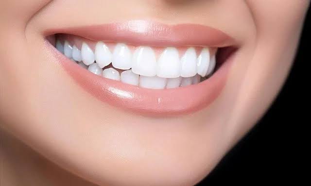
Once the clinical processes of restorative procedures applied in the field of oral and dental health are completed, long-term monitoring of the visual and functional gains achieved is highlighted as a medical necessity. Human anatomy does not have a static structure; bone density, gum levels, and jaw movements exhibit a natural change over the years. The porcelain laminates, zirconium crowns, or composite additions placed within this biological dynamism are constantly exposed to acidic changes and mechanical forces in the oral environment. Monitoring the adaptation of the applied Aesthetic Dentistry procedures over time forms the basic motivation for periodic clinical examinations.
Although individuals show great care for their daily home care, the conditions of microscopic boundaries where the tooth and prosthetic material meet, and the status of gum pockets can only be assessed in a clinical setting with professional tools. The monitoring strategies adopted in the Avrupadent clinical protocols are designed to support patients’ comfort after treatment and to detect material fatigue before it reaches significant levels.
How do clinical check-ups affect the durability of aesthetic procedures?
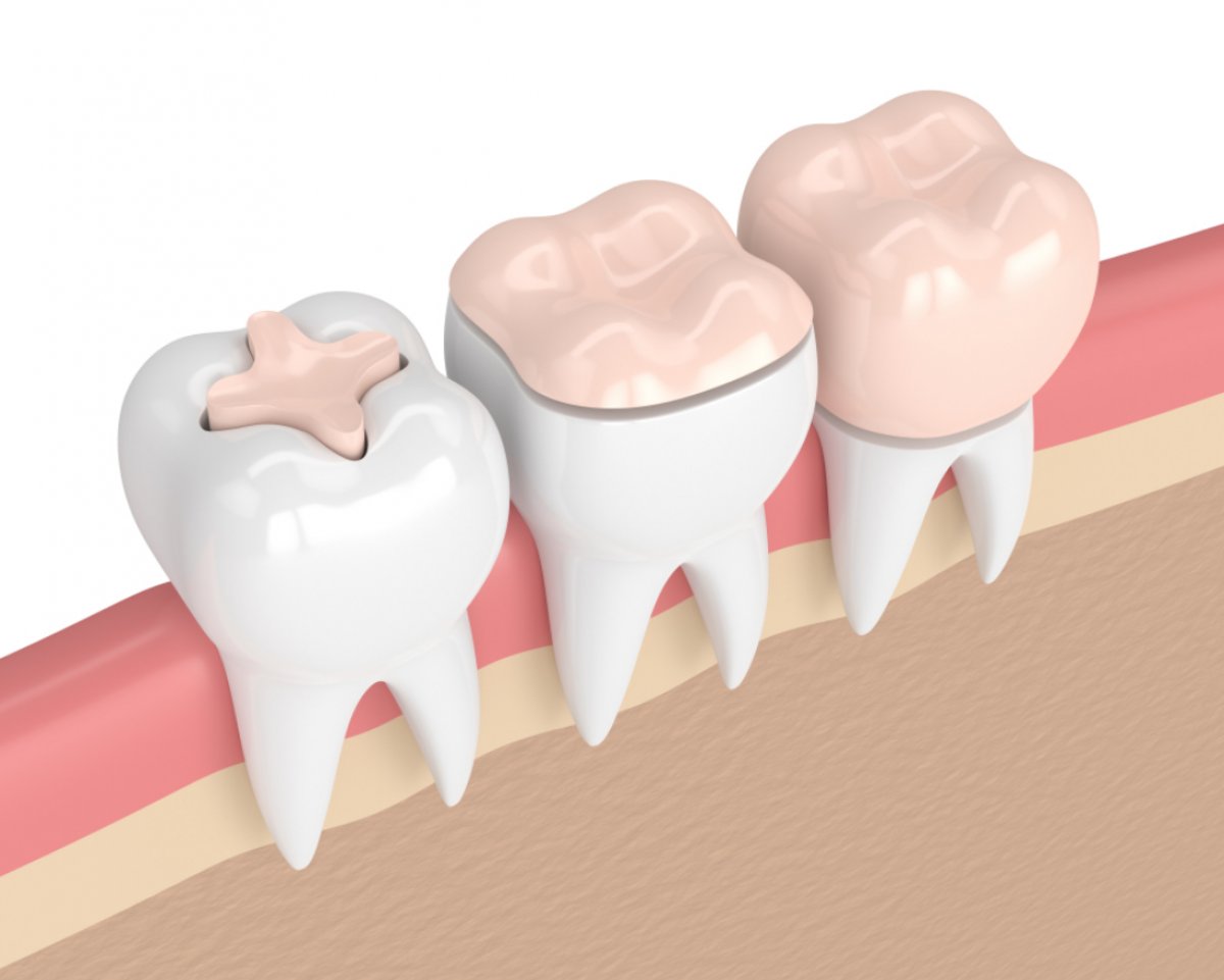
The porcelain, glass-ceramic, or nano-hybrid composite materials used in restorative dentistry are produced according to a specific breakage resistance in a laboratory environment. However, the oral environment is filled with continuously changing thermal variations (such as drinking cold water immediately after consuming hot coffee) and acid attacks. These thermal and chemical cycles can lead to microscopic signs of fatigue in the surface structure of restorations over years. Regularly conducted clinical examinations allow for monitoring the responses of these materials to environmental factors.
Especially resin-based materials applied directly, such as composite bonding, are more prone to water absorption and attachment of external pigments compared to porcelain. During routine checks, dentists re-establish the surface using special polishing rubbers and pastes to address any dullness on these materials. Keeping the surface free of plaque accumulation positively affects the material’s durability. Disruption of clinical follow-up processes poses the risk of these small deformations eventually leading to significant piece fractures.
| Material Type | Changes That May Occur Over Time | Intervention in Clinical Check |
|---|---|---|
| Porcelain Veneer | Dullness related to brushing in the surface layer. | Support of the surface shine with professional rubbers. |
| Zirconium Crown | The edge of the cavity in the tooth should be accessed. | Examination of tissue compatibility and updating of hygiene training. |
| Composite Bonding | Color tone deviations related to food pigments. | Re-polishing in a clinical setting. |
Why are Occlusal Changes Examined in Control Appointments?
The anatomy of the mouth is not a static structure; teeth engage in slow and microscopic movements throughout a lifetime. This natural process can lead to variations in the occlusal relationships of the upper and lower teeth over the years. A porcelain or zirconium crown placed for aesthetic purposes, even if it fits the occlusion perfectly on the first day, may face increased chewing forces later on due to microscopic positional changes in the natural teeth that occur over months.
Intense and disproportionate biting pressure (early contact) on a specific tooth can push the porcelain’s fracture resistance to its limits, potentially leading to cracks or traumatic tissue reactions in the jawbone supporting the tooth. During periodic examinations every six months, the patient’s occlusal balance is tested using special carbon (occlusal) papers. Very slight adjustments made at the identified pressure points help alleviate the stress on the restoration, contributing to the regular functioning of the chewing mechanism.
How Often Should Professional Care for Porcelain and Composite Surfaces Be Performed?
Daily brushing and flossing performed at home are essential for maintaining the cleanliness of teeth and restorations. However, patients’ personal efforts may sometimes be insufficient to remove stubborn bacterial plaques (biofilm layers) that form on the enamel surface or minor areas of the prosthetic margins. Particularly in individuals with heavy consumption of tea, coffee, and tobacco, surface discolorations can be observed in the margin areas of the porcelains and on the surface of composite fillings.
Professional cleaning (prophylaxis) carried out during clinical periodic examinations removes these deposits without damaging tissues by using ultrasonic tools and air-water spray devices. Subsequently applied fluoride-containing or protective pastes support the original surface smoothness of the restorations. Aesthetic Dentistry treatments ensure that these maintenance sessions are not missed, allowing the optical vitality of the restorations to be preserved over time.
How is the Gum (Periodontal) Compatibility Process Evaluated During Examinations?
The quality of an aesthetic restoration lies not only in its physical form but also in the harmony it displays with the surrounding pink tissue (gum). In the process following prosthetic procedures, if the patient neglects to use their interdental brush or dental floss, the first response will come from the gum tissue. The accumulation of bacterial plaque at the edges of the restoration can trigger an inflammatory reaction in the gums, leading to bleeding and mild swelling.
During periodic check-ups, the doctor thoroughly examines the color, consistency, and tightness of the gums surrounding the tooth. In areas where bleeding sensitivity is detected, the patient is provided with hygiene education specific to that area (modified brushing techniques, etc.) to encourage the tissue to return to a healthy state. Since gum issues detected in the early stages can lead to bone loss (periodontitis) if not intervened, these clinical follow-ups serve to protect the fundamental support of the aesthetic structure.
How is the Use and Effectiveness of the Night Splint Monitored During Check-Ups?
In individuals with tooth clenching and grinding (bruxism) habits, the clear night splints applied after aesthetic treatments serve as a barrier protecting the restorations from mechanical trauma. However, like teeth, these splints are also constantly exposed to high forces. Over time, wear, thinning, or small perforations may occur on the surface of the splint.
During control sessions, the doctor asks the patient to bring their night splint with them. The wear patterns on the splint provide the doctor with objective data about the directions in which the patient moved their jaw while sleeping. If there is any deformation in the form of the splint, adjustments will be made or it will be renewed if necessary. Monitoring the effectiveness of the splint in this way helps to reduce the risk of breaking porcelain laminates or composite restorations.
How Is Secondary Caries Formation Detected in Restoration Borders?
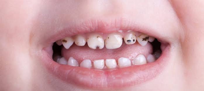
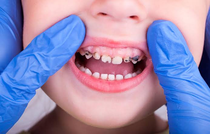
It is not expected for a tooth that has been crowned or filled to experience re-caries; however, the margin (border) areas where the restoration joins the natural tooth are biologically sensitive. Changes in saliva enzymes or acidic dietary habits can lead to weakening in the structure of adhesive cements over the years. Bacteria that leak from these weaknesses can initiate new lesions called “secondary caries” beneath the crown or at the edge of the filling.
The patient may not perceive these caries at the initial stage due to pain; since these lesions often progress very slowly. During periodic checks, the doctor mechanically tests all restoration margins using a dental probe (examination tool). Early detected micro-leaks can be resolved with small repair fillings without the need to remove the entire crown, while neglected cases may risk the liveliness of the tooth.
In Which Situations Are X-rays Required in Aesthetic Dentistry Procedures?
Visual clinical examination only provides information about the outer surfaces of the tissues inside the mouth. However, the root structure that forms the basic support of the tooth, periodontal attachments, and the jawbone lie beneath the gum tissue. The aesthetic compatibility of a porcelain crown depends on the health of the supporting bone structure of that tooth.
During certain stages of periodic checks (typically once a year or every two years), panoramic or periapical radiographs are taken to evaluate the internal adaptation of restorations and the tissues around the root (periapical tissues). This radiological monitoring supports the requirements of preventive medicine by allowing for the early detection of potential root tip infections or intraosseous cysts before symptoms are felt by the patient.
What Steps Are Followed for Control Appointments in Avrupadent Clinical Processes?
In corporate medical service approaches, the completion of treatment is not considered the end of the patient-doctor relationship, but rather the beginning of a sustainable protection period. Patients who have completed aesthetic processes in Avrupadent clinics are included in a follow-up appointment program tailored to individual tissue responses.
During the control sessions, assessments carried out by specialists in Aesthetic Dentistry encompass not only the examined teeth but an overall investigation of tissues within the entire oral cavity. Patients are motivated through the demonstration of the points they have neglected in home care, using anatomical models. This systematic process prepares the appropriate ground for the aesthetic and functional gains obtained to correspond to the patient’s quality of life for many years.
What are Frequently Asked Questions?
1. How often should I attend follow-up appointments after aesthetic procedures?
According to the principles of general dentistry, it is recommended to attend follow-up checks approximately every 6 months, although this may vary based on the individual’s oral hygiene and tissue responses.
2. Will I feel any pain during the follow-up examinations?
Follow-up sessions generally consist of visual examinations, occlusal tests, and surface polishing, which do not push physiological limits and are comfortable processes that do not cause pain for individuals.
3. How is the care of porcelain veneers conducted in the clinic?
Using special ultrasonic instruments and polishing (finishing) pastes that do not scratch the surface, plaques on and around the veneers are cleaned, and their original shine is maintained.
4. Should I bring my night guard to the follow-up appointments?
Yes, it is highly recommended to bring the night guard to appointments for testing its fit and for the doctor to examine the wear patterns on its surface.
5. Can composite fillings be renewed over time?
Composite resins may show slight dullness over time, but their surface brightness can be easily renewed with short-term polishing procedures applied during periodic check-ups.
6. How can it be understood that there is decay under the crown?
Dentists examine micro leakages by testing the crown margins with dental examination tools and evaluate the tissue beneath the crown with radiographic images when necessary.
7. Is taking an X-ray a requirement at every check-up appointment?
Taking an X-ray during every 6-month check-up is not mandatory; it can be planned annually or biennially based on the clinician’s clinical suspicion or to monitor the condition of the roots.
8. Is mild bleeding at the gum line a normal process?
Bleeding observed during brushing usually indicates plaque accumulation in the interdentals; this is not considered normal and should be supported with hygiene education during clinical check-ups.
9. Are zirconium crowns polished in the clinic?
Zirconium crowns are covered with baked porcelain in the laboratory; during check-ups, the attachments on this surface are cleaned to contribute to the continuity of its shiny appearance.
10. How is the use of dental floss monitored by the physician after aesthetic procedures?
The tightness and bleeding in the papillae (triangular tissues) at the gum level clinically demonstrate to the physician how effectively the patient has applied inter-dental cleaning (use of dental floss).
11. Do changes in chewing habits affect restorations?
Over time, changes in the jaw closure can occur due to unilateral chewing or external abrasions; this situation is balanced during check-ups as it may alter the load on restorations.
12. Can I go for check-up appointments during pregnancy?
Due to the hormonal changes that occur during pregnancy, it is recommended not to skip check-ups, but rather to maintain gum health with routine cleanings.
13. How long do clinical check-up sessions take?
Depending on the patient’s oral hygiene status and the number of restorations, a routine examination and polishing procedure generally takes between 20 and 30 minutes.
14. How is acidic beverage consumption detected during material checks?
Acid erosion can be identified by the doctor during surface examinations, as it can cause dullness and microscopic pitting in natural tooth enamel or composite surfaces.
15. How is the appointment schedule for Europadent organized?
At the end of treatment, a follow-up schedule is created based on the clinical needs of the patient, and the patient is informed in advance to ensure the process is managed in a compliant manner during upcoming examination periods.