

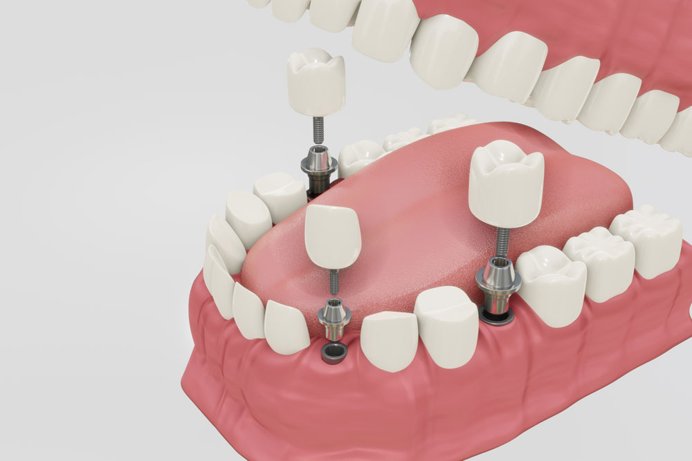
Will All Functions of the Tooth Return After Implant Treatment?
Tooth loss creates not only an aesthetic gap in human anatomy but also results in a physiological situation that directly affects chewing mechanics, the initial phase of the digestive system, speech articulation, and the cellular activity of the jawbone. Dental implants, which are part of the medical procedures applied to compensate for tooth deficiencies, are based on the principle of biocompatible titanium materials integrating with bone tissue. These clinical procedures, conducted in accordance with the Ministry of Health guidelines, aim to mechanically replace the lost anatomical structures. Avrupadent, operating in İzmir, addresses treatment planning with a multidisciplinary approach, aiming to restore the functional capacity of the patient in light of radiological and clinical findings.
How Does Implant Application Affect Chewing Function?
Titanium implants placed as replacements for missing teeth structurally integrate with the jawbone, directly transmitting chewing forces to the bone matrix. This mechanical connection contributes to the normal functioning of digestion by bringing patients’ biting and chewing capacity close to natural tooth physiology.
The process of chewing (mastication) in human anatomy is quite a complex mechanism that occurs when the lower and upper jaw teeth come into contact around a specific axis. During this action, the jaw muscles produce high amounts of vertical and horizontal forces to crush food. Natural teeth transfer these forces to the jawbone in a balanced manner through the periodontal connections (ligaments) around their roots. When tooth loss occurs, this mechanical transmission is disturbed; the person struggles to chew hard foods and develops a habit of unilateral chewing, placing asymmetrical load on the jaw joint (temporomandibular joint).
The function of implant-supported prostheses is to re-establish this broken biomechanical chain. When the titanium screw integrates completely with the bone at a cellular level (osseointegration), the chewing load acting on it is transferred directly to the underlying alveolar bone tissue, rather than to the mucosal (gum) surface as occurs with removable dentures. This structural feature is the fundamental medical element that distinguishes implants from traditional denture prostheses. In clinical practices based in Izmir, Avrupadent doctors determine the diameter and length of the implant to be applied according to radiological measurements based on the estimated chewing force (in Newton) that will act on that area. Since the rear molar teeth are the areas where the chewing force is felt most intensely, broad-diameter titanium roots are planned according to the anatomical limits of these regions.
Does Titanium Root Structure Affect Speech Articulation?
Tooth losses, particularly in the anterior region, can lead to issues with phonation (sound production) by altering the contact points of the tongue and the airflow inside the mouth. Preparing the anatomical forms of implant-supported prostheses to suit natural teeth allows the tongue and lips to adopt their normal position, facilitating the correction of articulation.
The act of speaking is formed as a result of the harmonious coordination between the air coming from the lungs and the vocal cords, palate, tongue, lips, and teeth. Particularly for the correct pronunciation of fricative (consonantal) and sibilant (hissing) consonants such as “s”, “f”, “v”, “z”, “t”, and “d”, the tongue must make contact with a specific pressure against the back surface of the upper front teeth or the incisive edges of the lips. In the absence of the upper front teeth, the airflow can escape uncontrollably outside the mouth, leading to speech impairments such as slurring or hissing sounds.
In cases where such deficiencies are attempted to be corrected with removable (customizable) prostheses, if the prosthesis covers the palatal area or makes millimeter adjustments inside the mouth, it can restrict the tongue’s movement area, affecting speech comfort. In implant applications, the artificial root is fixed to the natural bone, and ceramic crowns are placed in a laboratory environment to match the natural tooth contours. Since the roof of the mouth (palate) remains open, tongue movements are not restricted. In the prosthetic phase, the vertical dimension and horizontal overlap of the teeth are adjusted through clinical trials, restoring the pathways of airflow. Thus, the patient’s articulation ability is rebuilt within anatomical limits.
How Do Implant Teeth Arrange Jawbone Biomechanics?The jawbone requires chewing forces transmitted through the tooth roots to maintain its volume. The bone resorption process that begins after tooth loss can be controlled and slowed down thanks to the mechanical stress and cellular stimuli applied by the titanium implants placed in the region.
The jawbone requires chewing forces transmitted through the tooth roots to maintain its volume. The bone resorption process that begins after tooth loss can be controlled and slowed down thanks to the mechanical stress and cellular stimuli applied by the titanium implants placed in the region.
The relationship between mechanical loads on the human skeletal system and bone architecture is explained by “Wolff’s Law.” According to this law, bone tissue remodels itself based on the mechanical loads placed upon it. After tooth extraction, the region of the jawbone becomes functionless. The organism reduces mineral support to this unused tissue and osteoclastic (bone-resorbing) activity begins in the area. Volumetric losses occur in the bone tissue surrounding the extraction defect. In cases of prolonged tooth loss, the approximation of the jaw tip to the nose tip, the inward collapse of the lips, and the formation of an aging appearance on the face are the anatomical consequences of this bone resorption.
The titanium implant placed in the region through a surgical operation takes on the role of an artificial root when it achieves osseointegration (cellular bonding) with the bone. The chewing and biting forces applied to the prosthesis are transmitted to the bone through the titanium screw. This transmission mechanically stimulates the bone cells (osteoblasts) in the area, ensuring they remain active. In the processes of Avrupadent located in Izmir, the angle at which the implant will be placed, the distance to adjacent teeth, and the sinus cavities are calculated using tomographic measurements to ensure these biomechanical stimuli are transmitted in the correct axis. This medical procedure contributes structurally to protecting the existing bone volume.
| Physiological / Structural Features | Natural Teeth | Implant-Supported Teeth |
|---|---|---|
| Connection Shape to Bone | It is attached to the bone with flexible fibers called periodontal ligament. | Direct bonding of bone cells to the titanium surface (Osseointegration). |
| Force Absorption | Thanks to the flexible connections, part of the incoming force is absorbed by bending. | Since it is directly connected to the bone, there is no give, and it transmits force directly. |
| Touch and Pressure Sensation | It is very sensitive due to the nerve fibers around the root (proprioception). | Since there are no fibers around, sensation is limited to the inner bone nerves (osseoperception). |
| Infection Pathway | Gum inflammations are slowed down by periodontal connections before reaching the bone. | Due to the lack of connective tissue, infections caused by plaque accumulation can more rapidly affect the bone. |
How is the Functional Comparison of Implants and Other Methods in Missing Tooth Treatment?
Traditional dentures have a lower capacity to transmit chewing force compared to natural teeth because they are supported by the mucosa; on the other hand, bridge prostheses ensure chewing function but require the adjacent teeth to be reduced. Implants create stability close to natural physiology by transferring the force directly to the jawbone without intervening with adjacent teeth.
In clinical practice, there are three main prosthetic approaches used for the replacement of missing teeth: Removable dentures, fixed bridge restorations, and implant-supported prostheses. Removable (detachable) complete or partial dentures derive their support from the soft tissue of the gums and the existing bone underneath. This situation can lead to pressure on the mucosa during the consumption of hard foods, causing tissue irritation (trauma) and may result in loss of stabilization of the prosthesis. Chewing activities are biomechanically limited.
Fixed bridge prosthetics are restorations that use the healthy natural teeth on either side of the gap created by missing teeth as support (abutments). For the procedure to be applied, it is necessary to mill (prepare) the enamel tissues of these healthy teeth to a few millimeters. After the procedure, the chewing function is transferred to the supporting teeth through the bridge structure. Although this method offers a fixed use, the bone tissue in the toothless area under the bridge continues to resorb over time due to not receiving any mechanical stimulation.
Dental implant applications exhibit a more conservative (protective) approach to anatomy. No milling or abrasion is applied to the neighboring healthy dental tissues. Only a localized intervention is performed in the anatomical area where the tooth is missing. Since the force is transmitted directly from the artificial root to the alveolar bone, both biomechanical stimulation is provided and the integrity of the adjacent teeth is preserved.
How Does the Integration Process with Jawbone (Osseointegration) Determine Functions?
The osseointegration process, where titanium material integrates with the jawbone at a microscopic level, determines the capacity to bear the mechanical loads that will come onto the implant. In the lower jaw, this biological phase lasts an average of 2-3 months, and in the upper jaw, it lasts 3-6 months due to the difference in structural density; permanent prosthetic loading cannot be performed before this phase is completed.
The term osseointegration refers to a structural and functional connection in medical literature. The grooves of the titanium screw placed into the socket opened during the surgical procedure are invaded by bone-forming cells in the weeks following the operation. This phase, which begins with the organization of the clot, continues over time with the synthesis of new mineralized bone tissue. Since this bonding period is a cellular healing mechanism, it is medically impossible to shorten this period by any intervention applied from the outside.
Anatomically, the lower jaw (mandible) has a much denser, harder, and thicker cortical bone structure. This dense composition increases the mechanical stability (primary stability) at the moment the implant is first placed into the socket and accelerates the cells’ encapsulation of titanium. In contrast, the upper jaw (maxilla) has a more spongy (trabecular) bone architecture with adjacent sinus cavities containing air spaces. This structural difference leads to a longer anatomical spread of the healing and osseointegration phase in the upper jaw. In clinical procedures performed in İzmir, strictly adhering to waiting periods is the key to ensuring that the implant can resist chewing forces when porcelain or zirconium teeth are placed on it.
How is Implant Treatment Planning Carried Out at Avrupadent in İzmir?
The process at Avrupadent clinics involves: taking the patient’s systemic medical history, examining the bone anatomy with panoramic X-rays or three-dimensional tomography, performing the surgical procedure under local anesthesia, and after the recovery period, adapting functional upper prostheses into the oral cavity.
The clinical roadmap begins with a medical history (patient history) that primarily inquires about the patient’s overall physical health (conditions such as hypertension, diabetes, osteoporosis). Once it is confirmed that there are no systemic barriers to the procedure or that any chronic discomforts are under the control of a specialist physician, an intraoral examination is conducted. Digital imaging devices (CBCT) create a digital copy of the jaw. Critical anatomical reference points, such as the distance to the lower jaw nerve canal (nervus alveolaris inferior) and the height of the upper jaw sinus floor, are measured based on this digital data.
On the day of the operation, the preparation is carried out within the framework of asepsis and antisepsis (sterilization) rules suitable for surgical intervention in a clinical environment. Only the area to be operated on is anesthetized using local anesthetic agents, and nerve transmission is pharmacologically blocked. This allows the patient to maintain consciousness during the procedure, while still feeling mechanical pressure and vibration in the relevant area, the procedure continues within medical limits. After placing the titanium screw and suturing, postoperative care instructions are conveyed to the patient. When radiological confirmation is obtained that the biological waiting period has ended, the prosthetic phase begins, and the measurements and laboratory production of the teeth are completed.
- Systemic Evaluation: Blood sugar (HbA1c) levels, bone density, regularly used medications, and cardiovascular health are examined.
- Radiological Mapping: Three-dimensional tomography is used to perform millimetric analyses of the bone structure and create surgical guides.
- Surgical Stage: An incision is made in the gum under local anesthesia, sterilized saline solutions are used with micromotors to open the cavity and position the implant.
- Prosthetic Stage: After osseointegration, impressions are taken either with measuring materials or intraoral digital scanners and sent to the laboratory for the production of zirconium/porcelain crowns.
How Does Implant Superstructure Prosthesis Selection Shape Functional Capacity?
The prosthetic material (zirconium, metal-supported porcelain, monolithic ceramic) to be placed on the implant is determined based on the chewing area and the biting force of the patient. In the posterior regions, materials with pressure-resistant metal infrastructure are preferred, while in the anterior regions, zirconium restorations that provide natural tooth aesthetics through light permeability come to the fore.
Once the surgical process is successfully completed and the implant is firmly integrated with the bone, the functional outcome of the treatment depends on the choice of the prosthetic structure (crown, bridge). Different regions of the jaw are subjected to mechanically different chewing forces. In the molar (back teeth) region, chewing, grinding, and crushing actions exert significant vertical pressure. Therefore, the base of porcelain teeth to be used in these areas must be made of materials that are resistant to deformation and breakage, which is a clinical necessity. In this regard, metal-supported ceramics or specifically enhanced durable zirconium blocks are preferred.
In the anterior incisor regions, aesthetic expectations are prioritized alongside biomechanical needs. In restorations where an implant is placed in the front tooth area, it is desired that the reflection is from the functional tooth surface, and that the gum does not get influenced by the grayish color of the metal (not reflecting on the gum). The use of zirconium-based or all-ceramic E-max crowns is common in these areas. It is extremely important that the boundary of the gum (margin line) is accurately copied to the laboratory using digital or conventional measurements, so that the produced tooth functions in the mouth without causing food accumulation.
How Should Implant Care Be Done for Continuity of Function?
To maintain the long-term functionality of implant teeth, it is necessary to brush twice a day, ensure mechanical hygiene with intermediate brushes or mouthwash, and undergo clinical controls every 6 months. Insufficient hygiene can lead to infections known as peri-implantitis, resulting in bone tissue loss.
The acidic bacterial activities that cause tooth decay cannot structurally erode titanium, an inorganic metal, and the porcelain structure on it. However, the supporting tissues surrounding the implant, such as the gums (mucosa) and alveolar bone, are extremely sensitive to bacterial dental plaque. The periodontal ligaments surrounding the roots of natural teeth serve as a biological defensive wall that slows the progression of gum inflammation into the bone. Because this supportive tissue is not present around implants, the accumulated bacterial plaque (tartar) shows a tendency to rapidly seep into deeper tissues.
After treatment, oral care routines should go beyond just regular brushing. The underside of bridge abutments should be thoroughly cleaned using dental flossers (superfloss) or water flosser devices. Neglect can lead to inflammation beginning in the gums (peri-mucositis), which can evolve into a clinical picture known as “peri-implantitis” that erodes the bone around the implant if not treated. The particularly emphasized point by the doctors of Izmir Avrupadent to their patients is that the end of the prosthetic process is not a conclusion but rather the beginning of a long-term medical maintenance period, which includes regular oral hygiene and periodic dental tartar cleaning appointments. Individual mechanical cleaning and professional clinical check-ups are essential to maintain the stability of the achieved chewing and speaking functions.







