

Is the Implant Suitable for Everyone? Dental Treatment for Missing Teeth in Izmir
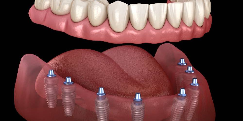
One of the frequently used biological restoration methods in the current medical literature for the replacement of missing teeth is the surgical placement of titanium alloy artificial roots into the jawbone. The mechanism of integration of this inorganic material at the cellular level with human physiology (osseointegration) requires the presence of certain anatomical and systemic conditions. Therefore, the answer to the question, “Is the implant suitable for everyone?” is examined on a wide range including the individual’s skeletal maturity, existing chronic discomforts, bone density, and parameters of the circulatory system. Within the 2026 guidelines of the Ministry of Health, decision-making processes for these types of surgical applications are carried out with objective medical analyses specific to the individual. Avrupadent, located in Izmir, determines individuals’ biological suitability by designing treatment protocols based on multidisciplinary clinical findings and three-dimensional radiological assessments.
Who Anatomically Can Undergo Implant Dental Treatment?
Anatomically, it can be applied to adult individuals who have completed skeletal development, possess certain width and height values in the jawbone, and have a general systemic health status suitable for surgical procedures. In patients without a medical contraindication, it is a medical procedure evaluated within clinical standards to replace missing teeth.
In jaw surgery practice, the anatomical structure of the operation area plays a determining role in terms of the long-term mechanical function of the titanium screw. The human jawbone consists of two different histological sections: cortical (hard outer surface) and trabecular (spongy inner structure). For a surgical cavitation (socket preparation) procedure to be performed, it is physiologically necessary to have at least 1.5 – 2 millimeters of additional bone wall thickness beyond the diameter of the implant to be used in the relevant area. The vertical length of the bone is limited by the distance to the mandibular nerve canal in the lower jaw and the maxillary sinus cavities in the upper jaw.
The presence of sufficient bone volume alone is not a sufficient parameter; at the same time, the thickness of the peri-implant mucosa (keratinized mucosa) covering this bone matrix is also desired to be of a specific bio-volume. A wider peri-implant mucosa acts as a protective soft tissue shield against microbial dental plaque that may accumulate around the prosthesis in the following months. In medical evaluations within the Europe-dent based in Izmir, both hard bone tissue and soft gum tissue are classified biotypically, and the suitability of the patient’s anatomical structure for this surgical procedure is mapped based on objective data.
Can Implants Be Placed in Individuals Who Have Not Completed the Development of Jawbone?
In individuals who are still in the growth and development phase (typically under the age of 18), implant surgery is not medically preferred due to the continuous changes in the size and volume of the jawbone. An artificial root placed at an early stage may remain in anatomically different positions, leading to asymmetric closures and orthodontic incompatibilities.
The skeletal components of the human body retain their active state until the end of puberty. The growth of the jaw-face (maxillofacial) region completes simultaneously with the ossification of growth plates (epiphyseal cartilages) in the wrist or cervical vertebrae. Natural teeth can move together with the jawbone via flexible tissues called periodontal ligaments and can continue the vertical slipping (eruption) movement.
However, inorganic titanium screws achieve ankylosis (fixation) by directly contacting bone cells. Because there are no periodontal ligaments around them, while the patient’s jaw continues to expand in both vertical and horizontal planes, the implant remains stable in the original bone coordinates where it was placed. As a result of this physiological difference, while natural teeth continue to grow over the years, the implant-supported restoration remains at a lower level. The clinical picture referred to as “infraocclusion” causes the opposing tooth in the arch to grow towards that space and disrupts the occlusal balance. Due to these biomechanical reasons, when tooth loss occurs in young individuals, space maintenance is achieved using temporary removable prostheses or orthodontic space maintainers until the growth period ends.
Do Systemic Diseases Inhibit Implant Application?Severe systemic diseases that are uncontrolled and in active phase (uncontrolled cardiovascular diseases, advanced immune deficiency) may limit surgical procedures medically. If systemic diseases are pharmacologically regulated under the supervision of a relevant specialist physician, the procedure is reassessed.
Severe systemic diseases that are uncontrolled and in active phase (uncontrolled cardiovascular diseases, advanced immune deficiency) may limit surgical procedures medically. If systemic diseases are pharmacologically regulated under the supervision of a relevant specialist physician, the procedure is reassessed.
Dental procedures, despite being localized surgical interventions, directly interact with the overall physiology of the organism. The patient’s medical history (anamnesis) is the primary factor determining whether the surgical intervention can be completed and the course of post-operative (after the procedure) wound healing. In medical literature, patients’ general health statuses are also evaluated according to the physical status classification of the American Society of Anesthesiologists (ASA).
Individuals classified as ASA I (healthy) and ASA II (having mild systemic disease, for instance, type 2 diabetes controlled by diet) can undergo clinical surgical procedures with standard medical protocols. However, for patients categorized as ASA III (severe systemic disease limiting functional capacity) or ASA IV (severe systemic disease posing a life threat), intensive medical consultations before the operation are essential. In the clinical processes of Avrupadent in Izmir, the pharmacological interactions of the medications used periodically by patients are analyzed, systemic blood tests are examined, and multidisciplinary approaches are followed by obtaining approval from relevant branch physicians in accordance with medical consent (waiver) forms.
| Type of Systemic Disease | Potential Physiological Effects on Surgery | Clinical Evaluation Approach |
|---|---|---|
| Uncontrolled Diabetes (Type 1 / Type 2) | Slows wound healing by disrupting microvascular (capillary) circulation, increases the tendency for infection. | HbA1c levels should be within a specific reference range. Prophylactic antibiotics may be started before surgery. |
| Cardiovascular Diseases | The use of anticoagulants can lead to prolonged bleeding in the surgical field, and may trigger stress hypertension. | A cardiology consultation is requested. Medication dosages are adjusted to be suitable for the operation, and anesthetics without adrenaline or with low adrenaline are chosen. |
| Osteoporosis | Intravenous use of bisphosphonate group medications poses a risk of jaw bone necrosis (osteonecrosis). | Consultation with an endocrinologist is necessary. The history of medication use (duration and dosage) is reviewed to shape the surgical schedule. |
| Immunosuppressive Conditions | A history of chemotherapy, radiotherapy, or organ transplantation affects white blood cell production and bone structure. | The approval of an oncology specialist is obtained. Surgical procedures for patients undergoing active treatment are planned for a later date. |
Can Diabetic Patients Undergo Implants?
If the blood sugar levels and glycosylated hemoglobin (HbA1c) values of diabetic patients are within reference ranges (generally around 7%), surgical procedures can be performed under infection control protocols. If there is uncontrolled high sugar, the procedure carries medical risks due to impaired cellular circulation.
Diabetes mellitus is a condition that results in hyperglycemia (high blood sugar) due to a metabolic defect in the body’s insulin production or utilization. Chronic hyperglycemia can lead to thickening and blockages in the walls of small blood vessels (microangiopathy) in the body. Oral tissues and the jawbone also suffer from this circulatory disorder. When a surgical incision is made and a cavity is formed in the bone, a strong blood flow is needed for the clotting mechanism and cell regeneration in that area. In the presence of uncontrolled diabetes, the transport of oxygen and leukocytes (defense cells) may be insufficient.
At the same time, high glucose levels disrupt collagen synthesis (tissue repair protein), slowing down the osteoblasts’ function of producing new bone. Opportunistic bacteria in the oral flora tend to multiply more easily in a high sugar environment. This situation increases the potential for infection around titanium screws. However, if the disease is regulated with a medical diet, oral antidiabetic agents, or insulin (under control), the intracellular biochemical reactions of these patients are quite similar to those of healthy individuals. In Avrupadent clinical protocols, it is aimed to ensure that fasting and postprandial blood sugar levels are measured on the morning of the operation day, remaining within the medical safety zone, and the surgical procedure is performed when these norms are maintained.
How Does the Implant Process Progress in Osteoporosis?
Although the diagnosis of osteoporosis alone does not create an absolute surgical limitation, the type of pharmacological agents used by the patient to slow down bone loss (oral or intravenous) and the duration of their use affect the clinical decision. Since these medications alter the healing mechanism of the jawbone, expert consultation is required.
Osteoporosis is a systemic skeletal disease characterized by a decrease in bone mineral density and a weakening of bone microarchitecture. This condition arises due to an imbalance between bone-forming osteoblast cells and bone-resorbing osteoclast cells, with the balance tipping in favor of resorption. One of the most commonly used drug groups in the treatment of osteoporosis is bisphosphonates, which inhibit the activity of osteoclasts to slow down bone loss. However, these drugs also slow down the natural remodeling cycle of the jawbone.
The administration of bisphosphonates in high doses over many years, especially via intravenous (IV) route, can lead to a specific clinical picture in jaw surgery. Following trauma to the site where surgery is performed, there is a risk of necrosis in that area of the bone due to halted remodeling cycles (Bisphosphonate-Induced Osteonecrosis of the Jaw – BRONJ). This medical complication occurs when the bone remains exposed and loses its vitality. In patients who use bisphosphonates in oral (tablet) form for a short duration, this risk is at much lower levels. In the İzmir Avrupadent environment, DEXA (bone density measurement) values and medication histories of osteoporotic patients are thoroughly examined, and it is decided in collaboration with endocrinologists whether to implement a drug holiday before the operation.
How Does Smoking Affect the Integration of Implants to Bone?
The nicotine and carbon monoxide found in tobacco products cause vasoconstriction in capillaries, limiting oxygen transport to the surgical area. This narrowing can hinder the integration process of the titanium screw into the bone matrix both mechanically and at the cellular level.
Smoking is one of the most significant external factors negatively affecting the biological healing functions of oral tissues and the jawbone. Carbon monoxide that enters the bloodstream through inhalation of cigarette smoke binds to hemoglobin molecules with a much stronger affinity than oxygen. This chemical reaction significantly reduces the transport of oxygen to tissues (tissue hypoxia). Angiogenesis, which is one of the initial stages of wound healing (the formation of new capillaries), is insufficient in an oxygen-deprived environment.
Additionally, the nicotine molecule reduces the volume of blood flow by constricting the vessel walls in the systemic circulation. Rich cellular nutrition is required for the blood clot formed around the implant cavity to organize into new bone cells (osteoblasts). Smoking also suppresses the production of natural protective immunoglobulin antibodies contained in tobacco, altering the bacterial profile in the oral cavity in favor of anaerobic (growing in oxygen-free environments) pathogens that lead to bone destruction. Clinical observations show that in heavy smokers, the surgical wound edges close less efficiently, and there are marginal losses observed in the gum (mucosal) tissue. Within the framework of medical requirements, it is recommended by physicians to limit smoking several weeks prior to surgical operation until the healing period is completed.
Is Implant Treatment Possible in Jawbone Insufficiency?
When further radiological examinations reveal that the jawbone is volumetrically insufficient, augmentation techniques such as bone grafting (bone powder) and sinus lifting are used to reconstruct the anatomy of the area. These additional procedures can prolong the healing period by adding specific recovery months to the medical treatment schedule.
In areas where the jaw has remained without stimulation for many years, bone resorption occurs due to a lack of biomechanical stimulation. This resorption narrows the width of the alveolar crest (the top of the bone) and shortens its vertical height. It is clinically necessary for the entire threaded surface of the implant to remain within the bone matrix to counteract the forces that will act upon it. If the screw extends beyond the bone, it can create a fenestration (windowing) or dehiscence (opening) beneath the soft tissues, resulting in an infection site.
To overcome these anatomical barriers, reconstructive surgical methods are employed in medical practice. Bone augmentation is performed using biomaterials in areas where the bone is thin. Graft materials serve as a cellular scaffold, allowing the body’s own bone cells to migrate into this structure and to harden. In the upper jaw, the maxillary sinus cavities, which are located above the roots of the upper teeth and assist with breathing, undergo expansion downward after tooth loss. In these cases, the surgical procedure (Sinus Lifting) of pushing the sinus membrane upward with special instruments and filling the resulting space with graft particles is performed. At the clinical stages of Izmir Avrupadent, the selection of graft and membrane (barrier) materials used during these medical operations is determined according to the three-dimensional anatomy of the patient’s bone defect.
| Material Classification | Source | Clinical Function and Biological Destruction Process |
|---|---|---|
| Autogenous Graft | Live bone blocks taken from the patient’s own jaw or outside the oral cavity. | Has the highest biocompatibility. The integration phase is organized as it contains live osteoblasts. |
| Xenograft | Animal-derived materials (mostly bovine or porcine) that are purified in a laboratory environment. | A calcium skeleton that contains no cells. It provides volume in the area for an extended period as it is slowly reabsorbed (dissolved). |
| Allograft | Particles that are obtained from human donors, sterilized and freeze-dried in tissue banks. | It stimulates bone induction, attracting surrounding cells to the area and transforming into the patient’s own bone. |
| Alloplastic Graft | Laboratory materials composed of synthetically produced calcium sulfate, hydroxyapatite, or bio-glass. | There is no risk of disease transmission. It creates inorganic substrates on which bone cells can grow. |
How is Implant Surgery Managed in Heart and Hypertension Patients?
In patients with cardiovascular disorders, the pharmacological agents used to thin the blood prior to the operation should be adjusted under the supervision of a cardiology specialist. In cases of hypertension, the vasoconstrictor components within local anesthetic solutions are selected specifically according to the patient’s current blood pressure parameters.
Cardiovascular health (cardiovascular system), during the medical management of oral surgeries, carries significant importance in terms of anesthesia selection, bleeding control, and stress management. In patients with coronary artery disease, heart valve prosthesis, or arrhythmia, the medications used—antithrombotic (aspirin-based) and anticoagulant (warfarin-based)—help prevent intravascular blockages by slowing down the coagulation cascade (mechanism). When an incision (cut) is made in dental practice, if the usual coagulation process does not occur, bleeding in the area cannot be controlled. Therefore, international coagulation measurement tests, known as INR (International Normalized Ratio), are examined. The use of blood thinners can be discontinued a few days before the operation in consultation with a cardiologist or may be transitioned to injectables with different molecular weights (bridging).
In individuals with hypertension (high blood pressure), the focal point is the selection of a local anesthetic agent. Among classic dental anesthetics, “epinephrine” (adrenaline) is included to extend the duration of effect and reduce bleeding in the surgical area. When adrenaline enters circulation, it increases the heart’s stroke volume and constricts blood vessels, which may lead to sudden spikes in blood pressure. To avoid this pharmacological effect in hypertensive patients, adrenaline-free or low-concentration anesthetic solutions are used. In the treatment processes in İzmir, Avrupadent practitioners adopt approaches that reduce clinical stress by also considering the natural adrenaline release triggered by dental phobia (fear) and monitor pulse and oxygen saturation with devices during the operation if necessary.
What Medical Criteria Determine Implant Planning at Avrupadent in İzmir?
Planning at Avrupadent clinics takes shape in line with 3D dental volumetric tomography measurements, the hospital’s detailed medical history, and analyses of alveolar bone biotype. Surgical stages are conducted with a multidisciplinary medical approach in accordance with national medical regulations, asepsis rules, and objective patient information standards.
The diagnosis phase, which is carried out before the application, is the most fundamental step that determines the course of the operation. Within clinical rotations, disciplines such as jaw surgery or periodontology examine the radiological data of the bone. Using a digital method called “backward planning,” the aesthetic and mechanical position of the zirconium or porcelain crown that will function in the mouth is designed in a computer environment. Subsequently, to ensure that the chewing forces are transmitted to the jawbone on the most balanced axis (vector) through this crown, the coordinates of the titanium screw in the bone are determined.
If necessary, these digital data are converted into surgical guide plates via 3D printers. During the operation, the use of these plates minimizes the possibility of damaging anatomical structures (nerves or sinus cavities) by limiting the entry point and depth of the drills (cutting tools). Surgical instruments are sterilized using vacuum steam sterilization in Class B autoclave devices, breaking the chain of infection. Located in Izmir, Avrupadent structures its treatment protocols not just based on marketing-focused expectations, but entirely on the anatomical realities of human tissue, cellular healing limits, and the patient rights regulations set by the Ministry of Health based on scientific standards.
What Should Mouth Care Be Like After Implant Application?“}For the long-term environmental stabilization of titanium material, specific hygiene routines should be established to prevent microbial plaque accumulation around prosthetic borders and in the oral mucosa (peri-implant mucosa). In addition to the use of brushing and interdental brushes, clinical dental calculus examinations should be carried out every six months.
For the long-term environmental stabilization of titanium material, specific hygiene routines should be established to prevent microbial plaque accumulation around prosthetic borders and in the oral mucosa (peri-implant mucosa). In addition to the use of brushing and interdental brushes, clinical dental calculus examinations should be carried out every six months.
Natural tooth enamel demineralizes and erodes when exposed to acid attacks due to calcium hydroxyapatite crystals in its structure. Titanium, being an inorganic and corrosion-resistant metal, does not pose a risk for dental caries. This anatomical difference can create a misconception among patients that the mentioned prosthetics do not require maintenance. However, the mucosal tissue that surrounds the implant surface and the underlying bone structure has a weaker defense mechanism against bacterial biofilm than natural teeth.
The periodontal fibers surrounding natural roots serve as a barrier that slows the progression of infection to the bone; however, this protective connective tissue is not present on the implant surface. Therefore, tartar (dental calculus) and bacterial plaque accumulating at the prosthetic borders (margins) can lead to inflammation in the soft tissue (peri-mucositis) and, if untreated, to the resorption of the jawbone (peri-implantitis). Among the care procedures communicated to patients by the experts at İzmir Avrupadent, in addition to tooth brushing, the mechanical cleaning of gaps under bridges with special threads called ‘superfloss’ or with water pressure dental devices is included. Ensuring radiological bone level controls during periodic clinical examinations conducted at least twice a year is a clinical necessity for the long-term functionality of the treatment.







